X-ray crystal structure and time-resolved spectroscopy of the blue carotenoid violerythrin
Oct 11, 2010
Oct 11, 2010
Prof John R Helliwell, University of Manchester
Violerythrin, a blue colored carotenoid, has been investigated by X-ray crystallography and by steady-state and ultrafast time-resolved absorption spectroscopy.
Carotenoids are isoprenoid polyene pigments that are widely distributed in nature. Besides their many functions, which include light-harvesting and photoprotection, an important biological role of carotenoids is natural coloration. Carotenoids are responsible for many of the vivid colors seen in bird feathers, tropical fish, crustaceans and flowers. The majority of carotenoids display yellow, orange or red color when isolated and dissolved in organic solvents, but a few rare examples of blue carotenoids exist.
The class of blue carotenoids may be divided into three groups. The first group consists of charged cation and oxonium ions of carotenoids, which can be obtained by treatment of the neutral molecules with Brønsted or Lewis acids. The second group represents carotenoid-protein complexes, in which non-covalent binding of a carotenoid to a protein causes a significant shift to longer wavelength of the absorption spectrum of the carotenoid compared to its spectrum in organic solvent.
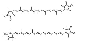 | |
| Figure 1. Molecular structures of s-cis- (top) and s-trans- (bottom) violerythrin. | |
This results in a blue color of the carotenoid-protein complex, despite the isolated carotenoid molecule having an orange or red appearance in organic solvent. For example, much work has sought to explain the large bathochromic shift of astaxanthin when it is bound in the protein β-crustacyanin, whose crystal structure has been determined by X-ray diffraction to 3.2 Å resolution. This structure allowed several possible molecular mechanisms responsible for the large bathochromic shift of free astaxanthin (absorption maximum at 475 nm in methanol) to astaxanthin bound to β-crustacyanin (absorption maximum at 570 nm) to be proposed, namely protonation of the conjugated carbonyl groups, hydrogen bonding to each of two histidine residues and two bound waters, excitonic interaction between the two astaxanthin molecules as well as planarization of the conjugated system. None of these effects alone appear to account for the entire bathochromic shift. The third group, of which violerythrin (Figure 1), the subject of this investigation, Diamond Light Source Annual Report 2010 is a typical representative, are neutral carotenoids that exhibit blue color even in organic solvents. Violerythrin contains a polyene chromophore (nonaene) with α-diketo groups on each of its terminal cyclopentene-dione end groups and exhibits blue color in organic solvents. Thus, violerythrin has four nonenolised conjugated carbonyl groups, a number unmatched by any other carbonyl carotenoid studied so far. In order to explore the controlling factors for blue coloration of neutral, carbonyl-containing carotenoids, we recently presented a comprehensive crystallographic and spectroscopic study of violerythrin in an international collaboration of investigators from several countries.
Dark blue needle crystals of violerythrin (Figure 2) were prepared by vapour diffusion techniques. Because the crystals were very weakly diffracting due to their small size, it was essential to carry out the data collection with intense monochromatic X-ray synchrotron radiation at the Diamond Light Source, Station I03, which is fed by an ultra-bright, undulator X-ray source. Using this data, the long awaited crystal structure of violerythrin (Figure 3 A,B) could finally be solved and refined. The crystal structure of violerythrin obtained at Diamond shows that the molecule is nearly planar with the terminal rings positioned in the s-trans conformation. This is in agreement with the NMR results, indicating that the solid state structure is the same as that in solution.
The steady-state and time-resolved spectroscopic data of violerythrin were found to be similar to those of other carbonyl carotenoids with long (N>10) π-electron conjugated chains. This indicates that while the four carbonyl groups in violerythrin are critical for generating the bathochromic shift that leads to the blue color of the molecule, no dramatic changes attributable to a charge transfer state, known to affect the excited state properties of carotenoids with short polyene chains, occur. This may be due to the symmetric distribution of the carbonyls, which would preclude such an effect. It should be noted that the UV/visible light absorption spectrum of the violerythrin in the solid state compares very well with the molecule in solution (Figure 4). This is obviously important in terms of being sure that the crystal structure represents the active coloration conformation.
Therefore it is concluded that the structural requirements for a blue, neutral, carotenoid are a planar, symmetric, cross-conjugated chromophore, containing at least thirty π-electrons, a central polyene chain with nine or ten conjugated carbon-carbon double bonds connected at each end by an s-trans or trans bond to two identical, cyclic end groups, each possessing a conjugated keto group further cross-conjugated to another keto group or a double bond in a quinoid type structure.
This research is at the interface of structural chemistry and structural biology and overall relates to the biological mechanisms of camouflage and/or photoreceptor light capture.
| |
| Figure 2 . A single crystal of violerythrin sitting in its loop-mount on Beamline I03 at Diamond; the crystal size is 0.12x0.04x0.02 mm3. |
References
[1] See Principal Publications and Authors (right).
[2] G.Britton and J.R. Helliwell ‘Carotenoid-Protein Interactions’ Chapter 6 in Carotenoids Volume 4: Natural Functions Edited by G. Britton, S. Liaaen-Jensen and H. Pfander Birkhauser Verlag, Basel-Boston-Berlin, 99-118 (2008).
[3] M. Helliwell ‘Three-dimensional Structures of Carotenoids by X-ray Crystallography’ Chapter 4 in Carotenoids Volume 4: Natural Functions Edited by G. Britton, S. Liaaen-Jensen and H. Pfander Birkhauser Verlag, Basel-Boston-Berlin, 37-52 (2008).
[4] Liaaen-Jensen, S.; Kildahl-Andersen, G. Arkivoc, 6, 5-25 (2008).
Principal Publications and Authors
Tomáš Polívka, Harry A. Frank, Miriam M. Enriquez, Dariusz M. Niedzwiedzki, Synnøve Liaaen-Jensen, Joanna Hemming, John R. Helliwell, and Madeleine Helliwell, X-ray Crystal Structure and Time-Resolved Spectroscopy of the Blue Carotenoid Violerythrin, J Phys Chem, Vol.114, Issue 26:8760-8769, (2010). DOI: 10.1021/jp101296a
Funding Acknowledgement
University of Manchester; National Science Foundation (MCB-0913022) and the University of Connecticut Research Foundation; Czech Ministry of Education (grants No.MSM6007665808, AV0Z50510513 and ME09037).
| Figure 3 | Figure 4 |
| |
| Figure 3. Plots of the violerythrin molecule; (A) ORTEP plot of the crystal structure using 50% probability ellipsoids; (B) plot viewed down the plane of the polyene chain, showing that the whole molecule is almost planar; hydrogen atoms have been removed for clarity. | Figure 4. Solid-state absorption spectrum of the crushed violerythrin crystals compared with the absorption spectrum of violerythrin in acetone solvent. |

Diamond Light Source is the UK's national synchrotron science facility, located at the Harwell Science and Innovation Campus in Oxfordshire.
Diamond Light Source Ltd
Diamond House
Harwell Science & Innovation Campus
Didcot
Oxfordshire
OX11 0DE
Copyright © Diamond Light Source. Diamond Light Source® and the Diamond logo are registered trademarks of Diamond Light Source Ltd
Registered in England and Wales at Diamond House, Harwell Science and Innovation Campus, Didcot, Oxfordshire, OX11 0DE, United Kingdom. Company number: 4375679. VAT number: 287 461 957. Economic Operators Registration and Identification (EORI) number: GB287461957003.