New structure helps explain so called children of the moon
Oct 7, 2010
Oct 7, 2010
Prof James Naismith, University of St Andrews
![]() Listen to Jim Naismith discuss this research in the Diamond podcast
Listen to Jim Naismith discuss this research in the Diamond podcast
DNA repair is found in all branches of life, it is the natural response to an environment which damages DNA. Defects in repair, structure of the archaeal protein XPD provides a model to interpret genetic mutations of the human homologue. These mutations lead to three related diseases: xeroderma pigmentosum (XP), trichothiodystrophy (TTD), and combined XP with Cockayne’s syndrome (XP/CS). The sequence homology allows us to use an orthologue from the organism Sulfolobus sulfataricus which we were able to solve the structure of. The structure shows that mutations can be grouped into two distinct sets, those that cause XP (& XP/CS) and those that cause TTD. The XP causing mutations affect directly the enzyme function, disrupting the helicase biochemistry. The role of these mutations has been confirmed biochemically. These mutations however preserve the function of the protein in forming the large repair machine. The TTD mutations preserve helicase activity but affect the ability of the protein to form the large multi protein complex. These mutations cluster on the surface areas and have been shown to preserve structure but affect stability of the large multiprotein complex.
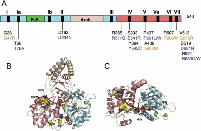 |
| Figure 1. Mutations of XPD leading to XP and XP/CS (A) Schematic showing the domain structure and canonical motifs of S. tokodaii XPD. (B) Structure of S. tokodaii XPD, with the 8-mer dU oligonucleotide derived from the co-crystal structure of the NS3 helicase shown in red. Residues mutated in XP and present in the S. tokodaii crystal structure are shown as space-filled sphere models, coloured by element (carbon, yellow; nitrogen, blue; oxygen, red). Residues R437 and A438 constitute part of the loop formed by motif V that is not observed in the crystal structure. Residue D182, in motif II (the Walker B box) is omitted for clarity.(C) Details as for (B), with the structure rotated vertically forward by 90°. (Reproduced from publication with permission). |
Human DNA is constantly being damaged by natural chemicals, sunlight and background radiation. Since these factors have all been around since the beginning of life, it is unsurprising that biology has evolved numerous mechanisms to repair DNA damage. The complete inability to repair DNA would be fatal to human life. However in humans incomplete or mis repaired DNA has serious health consequences, including cancer, premature aging and significant other illness. The repair machinery is multi protein and varies depending on the type damage. Such large complex machines are hard to analyse, by studying archaea which share a very similar (but simpler) repair system we can understand the human system. As part of one repair machine, the human helicase XPD unwinds DNA, mutations in XPD result in three related diseases: xeroderma pigmentosum (XP), trichothiodystrophy (TTD), and combined XP with Cockayne’s syndrome (XP/ CS). XP sufferers must avoid UV light as they cannot repair the damage and consequently are often seen after sun down, hence the popular name “children of the moon”. All three conditions are very serious often leading to early death, cancer and development issues.
|
| Figure 2. Mutations of XPD leading to TTD. (A) Schematic showing the domain structure and canonical motifs of S. tokodaii XPD. The positions of residues targeted by mutations causing TTD in human are indicated. S. tokodaii residues and numbering are in black and the equivalent human residue numbering indicated immediately below in red.(B) Residues targeted by TTD-causing mutations are represented in space-filled and labeled sphere models. Residue K84 is on the boundary of the FeS domain and is not visible in the crystal structure. (C) The model is coloured as in 2B and rotated 90° with respect to 2B to emphasize the bottom of domain 2. TTD mutations mapping to the bottom surface of motor domain 2 are coloured blue and labeled. In eukaryotic XPD, these residues together with the C-terminal extension probably form an interaction surface with the p44 protein.(D) Zoomed-in view of the boxed region of the Arch domain in Figure 2B. Residue A206 and surrounding residues are shown as space-filled molecular representations, with A206 in purple and other residues coloured according to their atom type. The TTD mutation introducing a tyrosine at this position (C259Y) is likely to cause significant disruption to the core of the Arch domain.(E) Plot showing the temperature stability of the wild-type and A204Y mutant XPD enzymes from S. acidocaldarius (equivalent to S. tokodaii A206Y and human C259Y). The helicase activity of the A204Y mutant is similar to that of the wild-type protein, but the mutant enzyme was inactive (Reproduced from publication with permission) |
The protein XPD is a FeS containing helicase that belongs to a wider superfamily including FancJ. Sulfolobus tokodaii is an archaeon that contains a close homologue of human XPD. Like the human enzyme, S. tokodaii is a 5’ to 3’ SF2 family helicase. Unlike the human protein it is readily tractable to structural study. We determined the structure of the protein to 2.25 Å using data collected at Diamond Beamline I03 in two passes ensuring maximum completeness at low resolution.
The crystal structure shows XPD contains the classic two motor domains (coloured salmon and cyan in Figures) that are thought to hydrolyse ATP during turnover. The protein also contains an Arch domain (coloured wheat) and the FeS domain (coloured green).
Sequence alignment of the human mutations onto the crystal structures, reveals very clear pattern. Figure 1 shows the XP (& XP/CS) mutations. These are clustered in the motor domain and would clearly affect the ability of the protein to carry out the helicase activity by disrupting the catalytic machinery and DNA recognition.
Figure 2 shows the TTD mutations. These are all remote from the catalytic site and are likely to affect protein protein interactions with perturbing catalysis. One of the mutants destabilises the structure of XPD but does not directly affect catalysis.
Principal Publication and Authors
Liu, L., Johnson, K.A, Rudolf, J., McRobbie, A-M., McMahon, S.A., Oke, M., Carter, L.G., Naismith, J.H. & White, M.F. Structure of the DNA repair helicase XPD. Cell, 133, 801-812 (2008).
Funding Acknowledgement
Biotechnology and Biological Sciences Research Council, UK Research carried out at Diamond on I03 and ESRF BM14.

Diamond Light Source is the UK's national synchrotron science facility, located at the Harwell Science and Innovation Campus in Oxfordshire.
Diamond Light Source Ltd
Diamond House
Harwell Science & Innovation Campus
Didcot
Oxfordshire
OX11 0DE
Copyright © Diamond Light Source. Diamond Light Source® and the Diamond logo are registered trademarks of Diamond Light Source Ltd
Registered in England and Wales at Diamond House, Harwell Science and Innovation Campus, Didcot, Oxfordshire, OX11 0DE, United Kingdom. Company number: 4375679. VAT number: 287 461 957. Economic Operators Registration and Identification (EORI) number: GB287461957003.