Macromolecular crystallography (MX) is a key technique in the structural biologist’s toolkit for understanding the function of biologically relevant molecules by revealing their shape and interactions at atomic resolution. The information derived from MX experiments can be complemented by many other techniques at Diamond for life science research (see the Soft Condensed Matter and Imaging and Microscopy sections of this review), coupled with experiments in the researcher’s lab, to giver deeper insight adopting an integrated structural biology approach.
At Diamond, seven beamlines (I03, I04, I04-1, I23, I24, VMXi, and VMXm), alongside the XChem fragment screening facility, the UK XFEL Hub, and the Membrane Protein Facility, are dedicated to exploiting the technique of MX for the benefit of the UK structural biology community, alongside researchers from Europe and further afield.
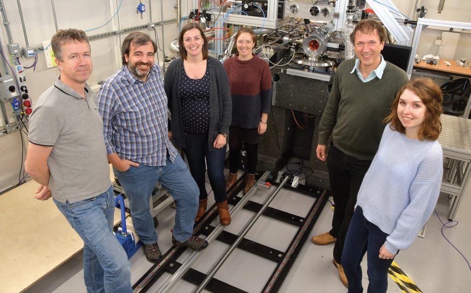
- First users and beamline team on VMXm, from left to right: Gwyndaf Evans (PBS), Jose Trincao, Anna Warren, Emma Beale, Diamond, and Ivo Tews and Rachel Bolton, University of Southampton.
It has been an exciting year for the VMXm micro/nanofocus MX beamline. After performing the first protein diffraction experiments at room temperature and in vacuum back in May 2018, first user experiments followed in October 2018. The group of Dr Ivo Tews from University of Southampton measured data from their sample crystals mounted on cryoelectron microscopy grids held at cryogenic temperatures in the evacuated sample environment. The compact sample space incorporates an on-axis video microscope and a Scanning Electron Microscope (SEM) for sample visualisation. The SEM has already proved invaluable in identifying the location, size, and shape of micron sized crystals. The smallest X-ray beam size measured to date on VMXm is currently 0.4 x 1.2 μm, and further commissioning and optimisation will take place throughout 2019.
Beamline VMXi provides a highly intense X-ray beam for the study of crystals at room temperature, particularly in the media in which the crystals are grown. Through 2018, significant upgrades have been made that enable VMXi to progress to a full user programme, with the beamline being the test bed for the first 2nd generation Eiger2 X detector (4M), which is capable of collecting data at very fast rates with extremely high count rates - perfectly matched to room temperature data collection with a very intense X-ray source. A new sample viewing system, and improved alignment configuration, now enable accurate automatic collection of data sets from many 10s, if not 100s of crystals per hour. Early experiments with serial crystallography delivery methods have been trialled and will add a further strength to the beamline capabilities in the coming years.
In the push to increase throughput across the beamlines, upgrades and methods are continuously being rolled out across the facilities. One such development in the last year is a significant improvement in grid scans – the ability to raster a sample in the X-ray beam and collect data. Fast grid scans have been developed by beamline I24, and are already rolled out to the beamline as well as I03 and I04. Building on the approach used on I24 for fixed-target serial crystallography, samples can now be moved continuously during grid scans allowing data collection at the maximum frame-rate of the detector, with no compromise on positional accuracy. With grid scans now running at >100 Hz, many more samples can be rastered over during a shift, and the throughput of automated X-ray centering will significantly increase. Data collection using this approach on the hard X-ray Imaging and Coherence beamline I13 has reached rates of 600 Hz, with a positional accuracy of 50 nm, illustrating that detectors are now the limiting step in raster scanning.
- Katherine McAuley (PBS), Neil Paterson and Mark Williams on I03.
Building on the work for Eiger detectors on VMXi, the first of the Eiger2 X 16M series detector model has been installed on beamline I04 recently. It replaces the first generation Pilatus2 detector, and provides a significant number of advantages and improvements that are benefitting the user community and science output. Data acquisition rates have increased five-fold over the previous detector, and a typical data set can now be collected in less than 15 seconds. The Eiger2 detector’s much smaller pixel size, coupled with no read-out time and vastly increased count rate capability, is leading to high dynamic range data with reduced background noise, improving data quality, and is beneficial in resolving large unit cells. Recently we installed the second Eiger2 X 16M detector on beamline I03 to complement the MX beamline suite’s capabilities.
Remote data collections are routine on the cryo-MX beamlines at Diamond and, over the last few years, there has been a steady increase in the numbers of samples shipped to Diamond for remote sessions, with more recently up to 90 dewars (or 10,000 sample mounts) on-site at any one time. Consequently, the logistics of managing users’ dewars and pucks has become more complicated, requiring new systems for tracking and streamlining the process of setting up for remote experiments. Tracking has been resolved by barcoding pucks and shipping dewars, and utilising the experiment database ISPyB. In this way, every experiment is tracked, and the responsible Diamond scientist is automatically informed which dewars are needed and where they are. The puck barcodes are scanned as they are loaded into the robot storage dewar and, consequently, the user is presented with a list of their samples when they start the data acquisition software. All these tools will be essential over the next few months as sample throughput is likely to increase further following the installation of new, faster detectors and, when automated, queued data collections become more widely used.
In the push to increase throughput across the beamlines, upgrades and methods are continuously being rolled out across the facilities.

- The I04-1 beamline team, clockwise from left: Jose Brandao-Neto, Richard Gillams, Frank von Delft (PBS), Alex Dias, Romain Talon, Ailsa Powell, Alice Douangamath, Anthony Aimon, Rachel Skyner.
Automated collection of data on beamline I04-1 has underlined the success of the crystal-based fragment screening facility, XChem, which to date has supported more than 130 new screens from academia and industry for fragment-based drug discovery. In October 2018, the I04-1/XChem team hit the milestone of the 100,000th XChem crystal collection – just three and half years since the XChem programme started. To meet the MX user’s increasing demands for XChem, the platform has been expanded, and a dedicated XChem support team has been put in place. More information on the XChem facility can be found in the Integrated Facilities section.
The team of beamline I23 has been working to establish MX in a wavelength range which hasn’t been accessible before, and has required building the first MX beamline operating in vacuum. This has necessitated a completely new approach to sample delivery being developed. A variety of studies have already been published, mainly for experimental native phasing, however, one recent highlight has been the study of potassium binding in the selectivity filter for potassium channels (Nature Communications, DOI 10.1038/s41467-018-06957-w), where access to the potassium K-edge at I23 unequivocally identified the biologically relevant state without recourse to complex substitution experiments. I23 can access the absorption edges of calcium, potassium and chlorine allowing unambiguous identification of the nature of these atoms even at low resolution. Over the next few years we will be able to give further insight into binding of these important elements in biology, information which remains elusive in cryo-EM. In the coming months, significant improvements to the beamline will be made to facilitate user mode and, to optimise best use of time on I23, adaptors for the I23 sample holders have been developed that enable fast sample screening on beamline I03.
Serial crystallography experiments at MX beamlines are complemented by Diamond hosting the UK XFEL Hub, a Wellcome funded initiative. It has a remit to develop hardware and software for Serial Femtosecond Crystallography (SFX), alongside supporting user training and access to SPB/SFX (single particles, clusters and biomolecules SFX) at the European XFEL and other hard X-ray facilities worldwide. Recently the XFEL Hub was internationally reviewed, with strong backing of the work and research the Hub is doing, and its support of the UK life science community who are awarded XFEL beamtime around the world. More details on the XFEL Hub can be found in the Integrated Facilities section.

Diamond Light Source is the UK's national synchrotron science facility, located at the Harwell Science and Innovation Campus in Oxfordshire.
Diamond Light Source Ltd
Diamond House
Harwell Science & Innovation Campus
Didcot
Oxfordshire
OX11 0DE
Copyright © Diamond Light Source. Diamond Light Source® and the Diamond logo are registered trademarks of Diamond Light Source Ltd
Registered in England and Wales at Diamond House, Harwell Science and Innovation Campus, Didcot, Oxfordshire, OX11 0DE, United Kingdom. Company number: 4375679. VAT number: 287 461 957. Economic Operators Registration and Identification (EORI) number: GB287461957003.