In its twelfth year of experiments, Diamond is now operating with 32 beamlines and 11 electron microscopes. The next year will see the completion of the latest phase of construction when the DIAD beamline welcomes first users. Of the electron microscopes, nine are cryo-electron microscopes specialising in life sciences, with two provided for industry use in partnership with Thermo Fisher Scientific, and make up eBIC (electron Bio-Imaging Centre). The two remaining microscopes dedicated to advanced materials research are supplied by Johnson Matthey and the University of Oxford. These microscopes form ePSIC (electron Physical Science Imaging Centre) and are operated under strategic collaboration agreements to provide for substantial dedicated peer reviewed user access. Both the eBIC and ePSIC centres are next to the Hard X-ray Nanoprobe beamline (I14). For academic research, Diamond instruments (beamlines and microscopes) are free at the point of access through peer review. For proprietary research, access can be secured through Diamond’s industry team.
Following a restructure in 2018, the instruments are organised into eight science groups as described below.
Electron Microscopes
| Microscope | Main Capabilities | Accelerating Voltages | Operational Status |
|---|---|---|---|
| Titan Krios I | Cryo-EM, Cryo-ET | 80, 120, 200, 300 kV | Operational since 2015 |
| Titan Krios II | Cryo-EM, Cryo-ET | 80, 120, 200, 300 kV | Operational since 2016 |
| Titan Krios III | Cryo-EM, Cryo-ET | 80, 120, 200, 300 kV | Operational since 2017 |
| Titan Krios IV | Cryo-EM, Cryo-ET | 80, 120, 200, 300 kV | Operational since 2017 |
| Titan Krios V | Cryo-EM, Cryo-ET | 80, 120, 200, 300 kV | Operational since 2018 |
| Talos Arctica | Cryo-EM, Cryo-ET | 200 kV | Operational since 2016 |
| Glacios | Cryo-EM, Cryo-ET | 200 kV | Installed, March 2019 |
| Scios | Cryo-SEM, Cryo-FIB | 3 to 30 kV | Operational since 2017 |
| Aquilos | Cryo-SEM, Cryo-FIB | 3 to 30 kV | Operational since 2019 |
| JEOL ARM200F | Atomic scale STEM imaging, EELS, EDX, electron diffraction | 80, 200 kV | Operational since 2017 |
| JEOL ARM300F | Atomic scale TEM and STEM imaging, electron diffraction, 4D-STEM, EDX | 30, 60, 80, 160, 200, 300 kV | Operational since 2017 |
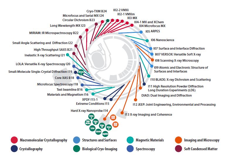
Diamond’s beamlines: current operational status April 2019
| Beamline Name and Number | Main Techniques | Energy / Wavelength Range | Status |
|---|---|---|---|
| I02-1 - Versatile MX micro (VMXm) | Micro- and nano-focus in vacuum cryo-macromolecular crystallography (VMXm) | 7 - 28 keV | Commissioning |
| I02-2 - Versatile MX in situ (VMXi) | In situ microfocus macromolecular crystallography, Serial Synchrotron Crystallography | 10 - 25 keV | Commissioning |
| I03 - MX | Macromolecular crystallography (MX), Multiwavelength Anomalous Diffraction (MAD) | 5 - 25 keV | Operational |
| I04 - Microfocus MX | MX, MAD | 6 - 18 keV | Operational |
| I04-1 - Monochromatic MX | MX, XChem fragment screening | 13.53 keV (fixed wavelength) | Operational |
| I05 - ARPES | Angle-Resolved PhotoEmission Spectroscopy (ARPES) and nano-ARPES | 18 - 240 eV; 500 eV | Operational |
| I06 - Nanoscience | X-ray Absorption Spectroscopy (XAS), X-ray photoemission microscopy and X-ray magnetic circular and linear dichroism | 80 - 2200 eV | Operational |
| I07 - Surface and Interface Diffraction | Surface X-ray diffraction, Grazing Incidence X-ray Diffraction (GIXD), Grazing Incidence Small Angle X-ray Scattering (GISAXS), X-ray Reflectivity (XRR) | 6 - 30 keV | Operational |
| B07 - VERSOX: Versatile Soft X-ray | Ambient Pressure XPS and NEXAFS | 250 - 2800 eV | Operational |
| NEXAFS and High-Throughput XPS | 50 - 2200 eV | Commissioning | |
| I08 - Scanning X-ray Microscopy | Scanning X-ray microscopy, NEXAFS/ XANES, X-ray fluorescence | I08 branch: 250 eV - 4.4 keV | Operational |
| J08 - Soft and Tender X-ray Ptychography branch: 250 - 2000 eV | Construction | ||
| I09 - Atomic and Electronic Structure of Surfaces and Interfaces | XPS (including HAXPES), X-ray Standing Waves (XSW), Near Edge X-ray Absorption Fine Structure (NEXAFS), Energy-scanned photoelectron diffraction | Hard X-rays: 2.1 - 18+ keV Soft X-rays: 0.1 - 2.1 keV (currently 0.1 - 1.9 keV) | Operational |
| I10 - BLADE: Beamline for Advanced Dichroism Experiments | Soft X-ray resonant scattering, XAS and X-ray magnetic circular and linear dichroism | Circular: 400-1600eV; Linear Horizontal: 250-1600eV; Linear Vertical: 480-1600eV | Operational |
| I11 - High Resolution Powder Diffraction | X-ray powder diffraction | 6 - 25(30) keV (0.5 - 2.1 Å) | Operational |
| DIAD: Dual Imaging and Diffraction | Simultaneous imaging and diffraction | 8 - 38 keV | Construction |
| I12 - JEEP: Joint Engineering, Environmental and Processing | Time-resolved imaging and tomography (phase- and attenuation-contrast), time-resolved powder diffraction, single crystal diffraction, diffuse scattering, energy dispersive X-ray diffraction (EDXD), high-energy small angle X-ray scattering (under development) | 53 - 150 keV monochromatic or continuous white beam | Operational |
| I13 - X-ray Imaging and Coherence | Phase contrast imaging, tomography, full-field microscopy (under commissioning), coherent diffraction and imaging (CXRD, CDI), ptychography and photocorrelation spectroscopy (XPCS) (under commissioning), innovative microscopy and imaging | Imaging branch: 8 - 30 keV | Operational |
| Coherence branch: 7 - 20 keV | |||
| I14 - Hard X-ray Nanoprobe | Scanning X-ray fluorescence, X-ray spectroscopy, ptychography and transmission diffraction | 5 - 23 keV | Optimisation |
| I15 - Extreme Conditions | Powder diffraction, single crystal diffraction | Monochromatic and focused 20 - 80 keV | Operational |
| I15-1 - XPDF | X-ray Pair Distribution Function (XPDF) | 40, 65, and 76 keV | Operational |
| I16 - Materials and Magnetism | Resonant and magnetic single crystal diffraction, fundamental X-ray physics | 2.5 - 15 keV | Operational |
| B16 - Test beamline | Diffraction, imaging and tomography, topography, reflectometry | 4 - 20 keV monochromatic focused 4 - 45 keV monochromatic unfocused White beam | Operational |
| I18 - Microfocus Spectroscopy | Micro XAS, micro Extended X-ray Absorption Fine Structure (EXAFS), micro fluorescence tomography, micro XRD | 2.05 - 20.5 keV | Operational |
| B18 - Core XAS | X-ray Absorption Spectroscopy (XAS) | 2.05 - 35 keV | Operational |
| I19 - Small-Molecule Single-Crystal Diffraction | Small-molecule single-crystal diffraction | 5 to 25 keV (0.5 to 2.5 Å) | Operational |
| I20 - LOLA: Versatile X-ray Spectroscopy | X-ray Absorption Spectroscopy (XAS), X-ray Emission Spectroscopy (XES) and Energy Dispersive EXAFS (EDE) | Dispersive branch: 6 - 26 keV | Optimisation |
| Scanning branch: 4 - 20 keV | Operational | ||
| I21 - Inelastic X-ray Scattering | Resonant Inelastic X-ray Scattering (RIXS), X-ray Absorption Spectroscopy (XAS) | Currently 250 - 1500 eV (to be upgraded to 250 - 3000 eV) | Optimisation |
| B21 - High Throughput SAXS | BioSAXS, solution state small angle X-ray scattering | 8 - 15 keV (set to 13.1 keV by default) | Operational |
| I22 - Small Angle Scattering and Diffraction | Small angle X-ray scattering and diffraction: SAXS, WAXS, USAXS, GISAXS. Micro-focus. | 7 - 20 keV | Operational |
| B22 - MIRIAM: Multimode InfraRed Imaging And Mircrospectroscopy | IR micro- & nano-spectroscopy, IR imaging, THz spectroscopy | nanoFTIR : 4000-900 cm-1 (2.5-11 μm) microFTIR: 10,000-100 cm-1 (1-100 μm) Spectroscopy (FTIR): 10,000-10 cm-1 (1-1000 μm) Imaging (FPA): 10,000-900 cm-1 (1-11 μm) | Operational |
| I23 - Long Wavelength MX | Long wavelength macromolecular crystallography | 3 - 8 keV (1.5 - 4.1 Å) | Optimisation |
| B23 - Circular Dichroism | Circular Dichroism (CD) | 125-500 nm & 165-650 nm for CD Imaging at 50 μm resolution, 96-cell High-Throughput CD (HTCD) and High-Pressure CD up to 200 MPa | Operational |
| I24 - Microfocus and Serial MX | Macromolecular crystallography, MAD, Serial Crystallography | 6.5 - 25.0 keV | Operational |
| B24 - Cryo Transmission X-ray Microscopy (TXM) | Full field X-ray imaging | 200 - 2600 eV | Optimisation |

Diamond Light Source is the UK's national synchrotron science facility, located at the Harwell Science and Innovation Campus in Oxfordshire.
Diamond Light Source Ltd
Diamond House
Harwell Science & Innovation Campus
Didcot
Oxfordshire
OX11 0DE
Copyright © Diamond Light Source. Diamond Light Source® and the Diamond logo are registered trademarks of Diamond Light Source Ltd
Registered in England and Wales at Diamond House, Harwell Science and Innovation Campus, Didcot, Oxfordshire, OX11 0DE, United Kingdom. Company number: 4375679. VAT number: 287 461 957. Economic Operators Registration and Identification (EORI) number: GB287461957003.