Biological imaging at Diamond now has a new home with the recent formation of the Biological Cryo-Imaging Group. This brings together dedicated facilities for X-ray, light and electron microscopy at Diamond. The bending magnet beamline B24 is the source of X-rays for the full field cryo-transmission X-ray microscope dedicated to biological X-ray imaging and the beamline has also established a cryo super resolution fluorescence microscopy facility which is a joint venture between Diamond and the University of Oxford. The Biological Cryo-Imaging Group is completed with the recently established national centre for cryo-electron microscopy at Diamond, eBIC (Electron Bio-Imaging Centre).
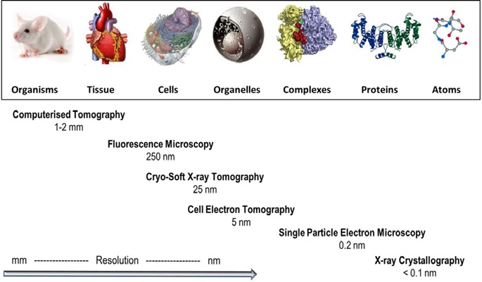
- Figure 1: Biological imaging in context. Representative techniques developed for the investigation of structure in biological systems at different scales, in order of resolution attainable. (Reproduced with permission from Harkiolaki et al. Emerg Top Life Sci (2018))

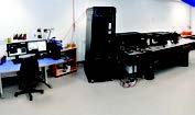
- Figure 2: B24 experimental hutch with the X-ray microscopy in the background.
B24 exploits cryo-soft X-ray tomography (cryo-SXT), which is a powerful technique for imaging intact cells in their near native state to resolutions of 25 - 40 nm. The technique sits neatly in the resolution gap that exists between electron and light microscopies and its real power lies in its ability to provide 3D imaging of whole cells with little or no chemical or mechanical modification. A schematic of the various imaging techniques available to biologists and how B24 and cryo-SXT fits into this tool set is shown in Figure 1. Cryo-SXT provides a unique tool to aid biologists to understand many key cellular and disease processes. One of the first external user experiments at B24 aptly illustrates the power of the technique where Hale et al used B24 to visualise red blood cells infected with the malaria parasite Plasmodium falciparum.
- Figure 3: Super resolution cryo fluorescence Microscopy facility at B24.
This work is summarised in more detail in the accompanying highlight. Sitting alongside the X-ray microscope at B24 is a cryo super resolution fluorescence microscopy facility, which is in the early stages of commissioning. It has also carried out first experiments with B24 users in the last year. Use of cryo-structured illumination microscopy (cryo-SIM) has allowed correlative imaging with the B24 X-ray microscope to be developed and workflows are currently being optimised which also paves the way for use with cryo-electron tomography (cryo-ET) at eBIC. The development of the facility is a joint collaboration with the advanced bioimaging unit, Micron, at the University of Oxford. B24 is now in the final round of commissioning and optimisation with users and will enter full user operations in the autumn of 2018.
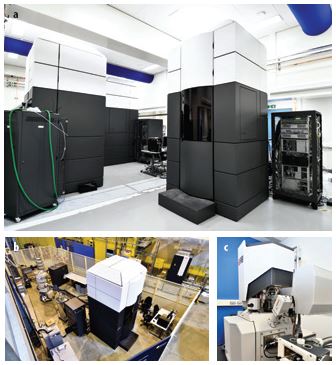
- Figure 4: (a) eBIC Krios hall housing Titan Krios II, III and IV microscopes; (b) Titan Krios I located next to I20 in the experimental hall of Diamond; (c) the SCIOS cryo focused ion beam scanning electron microscope at eBIC.
eBIC is the first high-end cryo-electron microscopy (cryo-EM) facility worldwide to be embedded in a synchrotron and user operations have been set up to mirror the well-established synchrotron beamline model. The centre is funded by the Wellcome Trust, the Medical Research Council (MRC) and the Biotechnology and Biological Sciences Research Council (BBSRC). eBIC has rapidly developed since it welcomed the first user group in July 2015. A key aim of the centre, enabled by the synchrotron access model, was to provide a state-of-the-art-facility for single particle cryo-EM and cryo-ET through cost-effective, peer-reviewed access based on scientific excellence. The rapid provision of high-end microscopes and the embedding within Diamond allowed this vision to be realised at breakneck speed and the success of the eBIC model has inspired other synchrotron sites worldwide to follow suit. In addition to providing cost-effective access to high-end microscopes for cryo-EM and cryo-ET, eBIC provides a focus for future hardware and software developments and advanced training for the community. The latter has been exemplified by specialised ‘hands-on’ training workshops in sample preparation for cryo-EM as well as specialised training courses in collaboration with the Collaborative Computational Project for electron cryo-microscopy (CCP-EM, ccpem.ac.uk) which is funded by the MRC and based at the Research Complex at Harwell. These courses and 'hands on' workshops have been extremely successful, although are heavily oversubscribed. This is due to sample preparation for cryo-EM remaining a major bottleneck and expertise in the community is lacking which is reflected in the greater than 10-fold oversubscription for the first two practical workshops dedicated to this topic at eBIC in January and October of 2017. Advanced training with support from CCP-EM, and the European funded Horizon 2020 project iNEXT (inext-eu.org), with which Diamond is a partner, will continue through 2018-19.
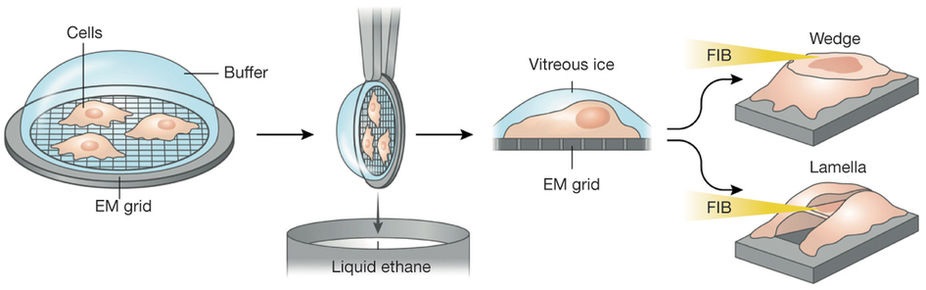
- Figure 5: Schematic of the workflow for cryo-FIB milling of cells with the SCIOS microscope at eBIC (Figure from Narayan K & Subramaniam S Nature Methods 12, 1021-1031 (2015)). Cells are grown on special grids for Transmission Electron Microscopy (TEM) and are rapidly plunge-cooled in liquid ethane and transferred to the SCIOS under cryogenic temperatures. A chosen area is FIB-milled tangentially either from one direction to produce a wedge or from above and below to produce a lamella, revealing the region to be imaged either using the scanning electron transmission mode in the SCIOS or it can be transferred to an eBIC Titan Krios under cryogenic conditions and imaged at high resolution using cryo-ET.
On the computing front, on-the-fly processing of data has been rolled out and management of data collected and processed at eBIC will be presented in the information system ISPyB that is now being developed for use by cryo-EM. The latter project is being coordinated by Diamond through the H2020 iNEXT project and will be available for use in the near future at all the European cryo-EM centres. These software developments have been carried out in collaboration with CCP-EM and the developers of the Scipion framework which brings together numerous software packages for EM and presents them to the user in a unified interface for both biologists and developers. This has enabled the accelerated development of these automated workflows at eBIC and ISPyB will provide added value and provides a key tool for users to allow remote access use of eBIC to be realised.
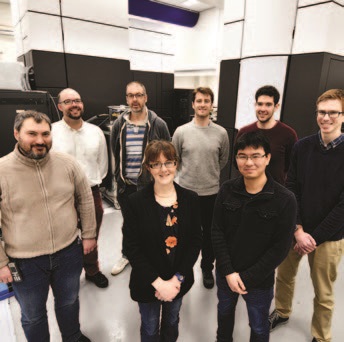
- Figure 6: Some members of the eBIC team in the Krios Hall.
The user program at eBIC has continued to grow rapidly in 2017 with two new Titan Krios 300 KeV microscopes (Thermofisher) brought online, taking the total number of high-end microscopes to 4. eBIC is also heavily engaged in developing a user program for cellular studies and during 2017 a cryo focused ion beam scanning electron microscope (SCIOS, Thermofisher) has been in commissioning with in-house users. The microscope allows the cryo-milling of cellular samples into thin slices (up to 100 nm) making them suitable for imaging by cryo-ET (a schematic of the workflow is illustrated in Fig. 4). Whereas X-rays are more penetrating and cryo-SXT at B24 can image samples as thick as 10-15 mm, cryo-ET requires thin cellular slices with the advantage that imaging at much higher resolutions are achievable (Fig. 1). Again, here a correlative approach that combines light and electron microscopy provides a powerful tool to image cellular regions of interest. In collaboration with B24 development of workflows that combine live-cell fluorescent light microscopy, cryo-fluorescent microscopy, and cryo-ET to provide a structural and dynamic view of cellular processes are in development. A call for proposals from external users for commissioning of the SCIOS microscope at eBIC was announced in April 2018 for experiments in early autumn 2018.
In summary the new Biological Cryo-Imaging Group at Diamond is at an exhilarating stage of development and we expect research and development to continue at a fast and exciting pace through 2019 as we bring all the instruments in the group fully online.

Diamond Light Source is the UK's national synchrotron science facility, located at the Harwell Science and Innovation Campus in Oxfordshire.
Diamond Light Source Ltd
Diamond House
Harwell Science & Innovation Campus
Didcot
Oxfordshire
OX11 0DE
Copyright © Diamond Light Source. Diamond Light Source® and the Diamond logo are registered trademarks of Diamond Light Source Ltd
Registered in England and Wales at Diamond House, Harwell Science and Innovation Campus, Didcot, Oxfordshire, OX11 0DE, United Kingdom. Company number: 4375679. VAT number: 287 461 957. Economic Operators Registration and Identification (EORI) number: GB287461957003.