Novel hybrid protocells may hint at origins of life

Many scenarios have been proposed for processes leading to the formation of the protocells that are a pre-requisite to life on early Earth. One theory is based on fatty acid vesicles, which form when naturally occurring fatty acids self-assemble in water to form a bilayer membrane. An alternative theory suggests that protocell formation may occur when organic chemicals and naturally occurring polymers present in aqueous solution spontaneously phase separate to form membrane-free chemically rich liquid microdroplets, called coacervates. Combining these two approaches, a hybrid protocell model has been developed based on the spontaneous self-assembly of a fatty acid membrane on coacervate microdroplets.

Beamline I22 Scientific Highlight

The most fundamental requirement for the emergence of cells on early Earth is the existence of a closed compartment, but how this came about remains a mystery. Among several possibilities, vesicles formed by the self-assembly of fatty acids have been the most intensely studied for origin of life studies1. Experiments on these and corresponding protocell models based on lipids and surfactants have demonstrated that these compartments are successful in spatially confining molecules and enzymes and undertaking localised catalysis and replication processes key to fundamental biochemical reactions2. Whilst these observations have provided strong support for the protocell vesicle, they are limited in that the membranes are often impermeable which restricts free exchange of molecules into and out of the cell and the vesicles do not mimic key cellular properties associated with molecular crowding. In addition, lipid synthesis is incredibly complex and it is plausible that simpler compartments were generated on the early Earth.
Membrane-free liquid droplets formed by coacervation offer an intriguing and alternative route for cellular organisation. Over 50 years ago, Oparin3 suggested that the spontaneous phase separation of counter-charged polyelectrolytes into chemically enriched compartments provided a facile route to prebiotic compartmentalisation, which was independent of membrane (vesicle) formation. There are a large number of examples of coacervation between biomolecules and macromolecules, and recent reports have shown that coacervate microdroplets can be formed from simple biological molecules such as low molecular weight peptides and mononucleotides, and ribonucelotides4. The resulting droplets are stable over a range of pHs and temperatures and consist of molecular crowded aqueous interiors, enriched in biomolecular components, which display enhanced catalytic activities5. The highly charged, polymer rich interior permits sequestration of a wide range of low and high molecular weight solutes including enzymes, substrates and photoactive molecules. The facile route to spatial compartmentalisation and chemical enrichment via coacervation is attractive, however, the lack of an enclosed membrane is a drawback of the model compared with vesicular protocells.

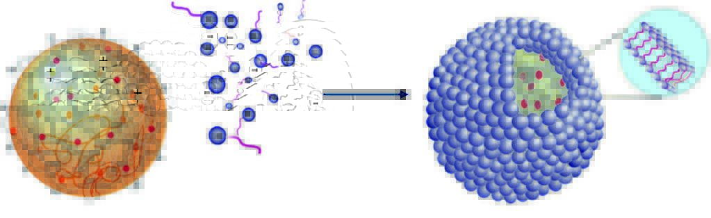
Figure 1: Schematic showing the self- assembly of fatty acid multilayers on the surface of a positively charged coacervate droplet.

To address the limitations of both models, a new type of protocell was designed and constructed, which integrates aspects of phase separated liquid droplets and fatty-acid vesicle self-assembly to produce a hybrid model of prebiotic organisation (Fig. 1). Simple physical and chemical processes were used to self-assemble continuous fatty acid multilayers on the surface of preformed coacervate microdroplets. Positively charged coacervate microdroplets of diameters of a few to several tens of micrometres, were prepared with binary combinations of short/long chain cationic peptides (oligolysine, polylysine) or a synthetic polyelectrolyte (polydiallydimethylammonium chloride) with negatively charged monoribonucleotide (adenosine triphosphate (ATP)) or ribonucleotides. Fatty acid monomers (oleic acid) were introduced to the dispersion of positively charged droplets as the oleate salt. Instead of selforganising into vesicles, due to the low concentrations, the negatively charged oleate monomers were attracted to the surface of the droplets. Further addition of oleate monomers led to the production of a continuous delineated, organic, membrane. When the coated and uncoated droplets were stained with a lipophilic tagged fluorescent dye (BODIPY), fluorescence optical microscopy images showed a transition from a homogeneous distribution of dye within uncoated droplets to a heterogeneous distribution with the coated droplets (Fig. 2). The increased fluorescence on the surface of the droplets indicated that there was an accumulation of fatty acid at the surface.

Small Angle X-ray Scattering (SAXS) experiments performed using the Small Angle Scattering and Diffraction beamline (I22) were then used in combination with Fluorescence Lifetime Imaging (Fig. 3a), to explore the microstructure of the coated coacervate droplets. The SAXS experiments showed that the fatty acid had assembled to form highly organised multilayers (Fig. 3b) localised on the surface of the coacervate (Fig. 3a). No multilamellar structures were seen in SAXS patterns at low fatty acid concentrations and light scattering experiments confirmed that little to no fatty acid vesicles were present in the dispersion (Fig. 3c). The very high intense X-rays available at Diamond coupled with I22’s extremely sensitive detector were necessary to determine the size and structure of the microcompartments with some key features of interest between 4 and 6 nm in size.

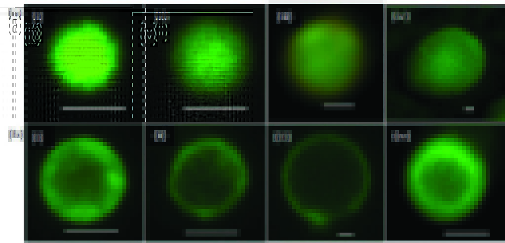
Figure 2: Fatty-acid membrane self-assembly on the surface of positively charged coacervate microdroplets. Fluorescence microscopy images of single droplets stained with the lipid-soluble BODIPY FL C16 dye. (a) Non-coated microdroplets showing homogeneous fluorescence throughout the interior of the microcompartments: (i) polylysine/RNA; (ii) oligolysine/RNA; (iii) oligolysine/ATP; (iv) PDDA/ATP. Scale bars, 1 μm. (b) Fatty acid coated droplets prepared from oleate/polylysine/RNA (i), oleate/oligolysine/ RNA (ii), oleate/oligolysine/ATP (iii) and oleate/ PDDA/ATP (iv) showing assembly of a fatty-acid membrane specifically at the surface of the microcompartments. Scale bars, 1 μm.

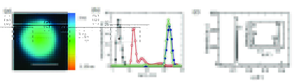
Figure 3: (a) Fluorescence lifetime imaging microscopy image of a fatty acid-coated coacervate droplet containing kiton red showing two different lifetimes and viscosities in the centre (coacervate matrix) and edge regions (lipid membrane); scale bar = 5 μm. (b) Small angle X-ray scattering profile showing Bragg reflections corresponding to a multilamellar fatty acid membrane at the surface of the coacervate droplets. (c) DLS profiles showing volume distributions of hydrodynamic diameters (DH (nm)) for oleate micelles (black filled squares), oleic acid/oleate multilamellar vesicles (red open squares), uncoated PDDA/ATP microdroplets (blue filled circles,) and oleate-coated PDDA/ATP microdroplets (green open circles).Caption with parts indicated where necessary; (a) first part; (b) second part.

The ability of the fatty acid membrane to establish concentration gradients across the membrane was tested by incubating the hybrid protocells in the presence of dyes with a range of molecular weights and charges. Fluorescence confocal microscopy images showed that the uptake properties of the droplets were altered in the presence of the membrane where positively charged dyes were generally excluded from the droplet interior compared to the membrane free systems. Finally, the internal polyelectrolyte/ ribonucleotide droplets could be disassembled under high ionic strength (75 mM NaCl) whilst retaining the oleate multilayer membrane to produce fatty acid vesicles that contained concentrated aqueous solution of encapsulated polyelectrolyte and ribonucleotide molecules.
The studies using SAXS were crucial in confirming the presence of multilayers within the aqueous dispersion and in combination with a range of techniques was able to conclusively show that a new type of hybrid protocell could be constructed using simple chemical and physical laws.

Source publication:
Tang, T. Y. D., Hak, C. R. C., Thompson, A. J., Kuimova, M. K., Williams, D. S., Perriman, A. W. & Mann, S. Fatty acid membrane assembly on coacervate microdroplets as a step towards a hybrid protocell model. Nature Chemistry 6, 527-533, doi:10.1038/nchem.1921 (2014).

References:
1. Monnard, P. A. & Deamer, D. W. Membrane self-assembly processes: Steps toward the first cellular life. Anatomical Record 268, 196-207, doi:10.1002/ ar.10154 (2002).

2. Oberholzer, T., Wick, R., Luisi, P. L. & Biebricher, C. K. Enzymatic rna replication in self-reproducing vesicles - an approach to a minimal cell. Biochemical and Biophysical Research Communications 207, 250-257, doi:10.1006/bbrc.1995.1180 (1995).

3. Oparin, A. I. The Origin of Life, 2nd ed. Dover Publications: Dover (1953).

4. Koga, S., Williams, D. S., Perriman, A. W. & Mann, S. Peptide-nucleotide microdroplets as a step towards a membrane-free protocell model. Nature Chemistry 3, 720-724, doi:10.1038/nchem.1110 (2011).

5. Crosby, J. et al. Stabilization and enhanced reactivity of actinorhodin polyketide synthase minimal complex in polymer-nucleotide coacervate droplets. Chemical Communications 48, 11832-11834, doi:10.1039/ c2cc36533b (2012).

Funding acknowledgements:
We thank the Engineering and Physical Sciences Research Council (EPSRC, UK) and European Research Council (Advanced Grant) for financial support. We thank the EPSRC for a Career Acceleration Fellowship (grant number EP/E038980/1) to M.K.K. and the Malaysian government for the award of a PhD studentship to C.R.C.H. We acknowledge beamline I22 (N. Terrill and A. Smith) at Diamond Light Source, W. Briscoe for beamtime and R. Richardson and M. Thomas for assistance and use of software for X-ray analysis.

Corresponding author:
Professor Stephen Mann, University of Bristol, s.mann@bristol.ac.uk

Diamond Light Source

Diamond Light Source is the UK's national synchrotron science facility, located at the Harwell Science and Innovation Campus in Oxfordshire.

Copyright © 2022 Diamond Light Source


Diamond Light Source Ltd
Diamond House
Harwell Science & Innovation Campus
Didcot
Oxfordshire
OX11 0DE

See on Google Maps

Diamond Light Source® and the Diamond logo are registered trademarks of Diamond Light Source Ltd

Registered in England and Wales at Diamond House, Harwell Science and Innovation Campus, Didcot, Oxfordshire, OX11 0DE, United Kingdom. Company number: 4375679. VAT number: 287 461 957. Economic Operators Registration and Identification (EORI) number: GB287461957003.

feedback