“These pili are important surface-exposed appendages that bacteria use to recognize and adhere to host tissues. They are also important in making bacterial biofilms,” said researcher Gabriel Waksman, PhD, of UCL and Birkbeck College London. “The main challenge is that, until recently, there was no method to determine the atomic-resolution details of these appendages. However, recently, revolutionary progress in electron microscopy has changed that. We can now generate views of these pili at very high resolution, yielding unprecedented atomic details that shed light into the function of these pili.”
Understanding the shape and structure of the pili is a key step toward producing ways to block the bacteria from setting up shop and producing the common and often painful infections. (It’s estimated that up to half of women develop a UTI during their lifetime.) Doctors might, for example, develop drugs that would prevent the pili from getting a toehold in the urinary tract. And that’s an exciting prospect.
“These pili are absolutely essential for the infectivity, because it’s the pili that attach very strongly to the lining of the urinary tract,” Egelman noted. “If these pili aren’t assembled, then these bugs aren’t infective at all. They’d wash right out.”
David Stuart is Director of Life Sciences at Diamond and Director of the eBIC facility. He comments: “It’s fantastic to see the tremendous potential of cryo-electron microscopes being exploited in this research. This technology is allowing us to explore complex biological systems in unprecedented detail, yielding groundbreaking findings such as this one. Ultimately, work such as this has the potential to support new drugs and therapies that will make all the difference to individual patients.”
The new research has been published online by the scientific journal Cell.
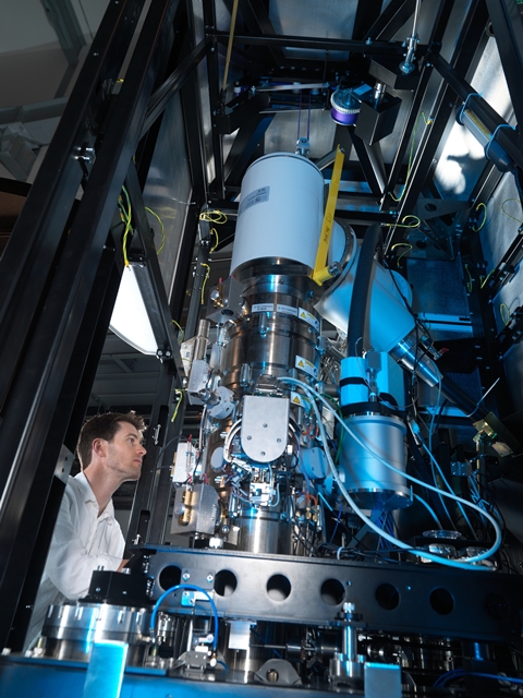
Alistair Siebert inspects the Krios electron microscope.