Scientists have used Diamond Light Source to advance our understanding of the changes taking place during the progression of brain cancer. This research may lead the way to a new tumour assessment method which could complement traditional approaches.
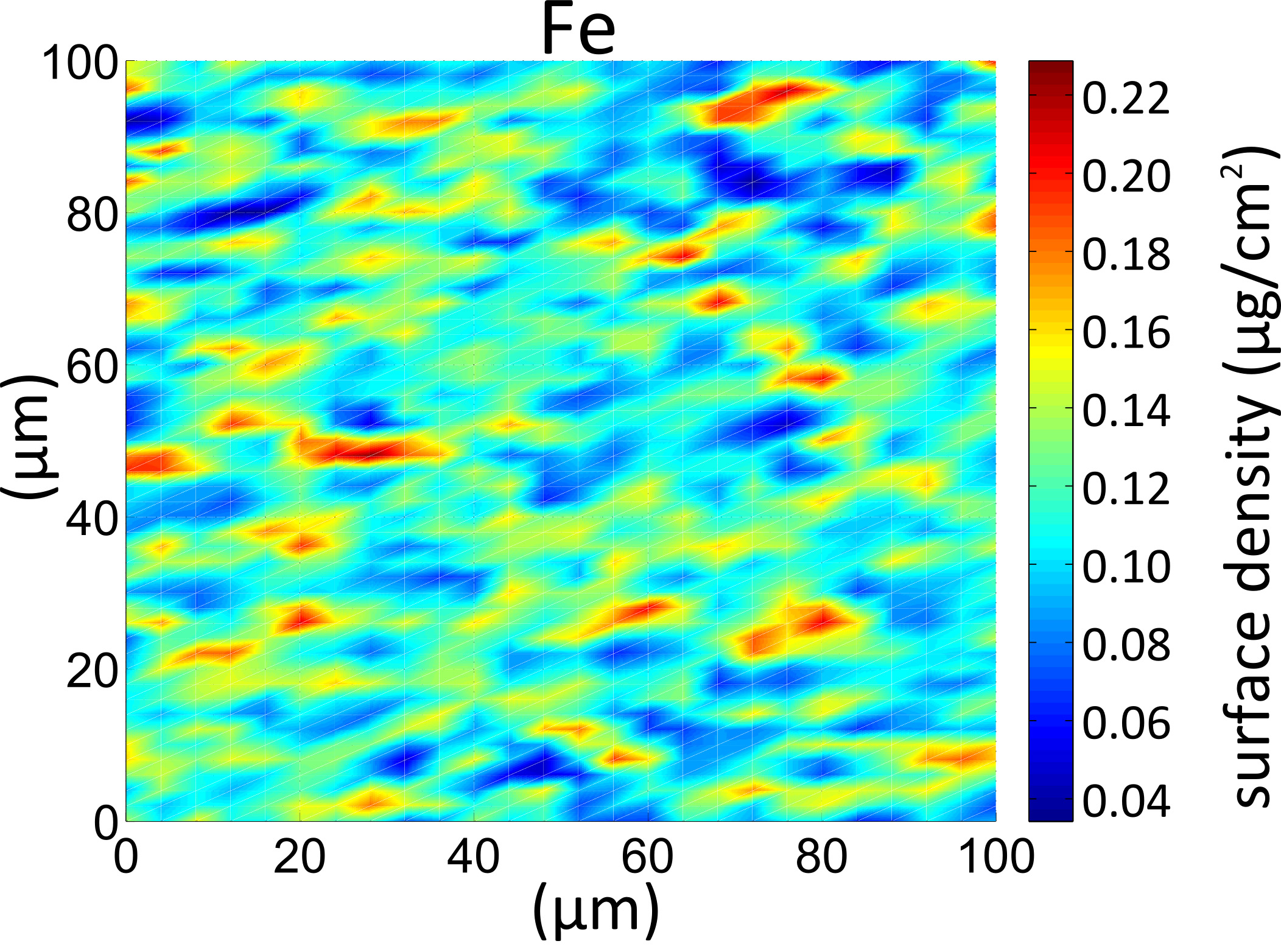
Changes in metal distribution in the brain have been linked to the degree of malignancy of brain cancer. With results published in Spectrochimica Acta Part B, the researchers found that trace metals could be used to correctly identify cancerous tissue in over 99% of cases and effectively classify the cancer stage.
The 4,000th paper to be published based on research carried out at Diamond, this work could have major implications for the early identification and treatment of brain tumours.
Diamond’s CEO, Andrew Harrison, comments: “I’m delighted that Diamond’s 4,000th paper so aptly demonstrates the impact that synchrotron research can have on people’s lives. This work is still in its early stages but, in time, the discovery of the link between certain trace metals and their role in the growth of cancer cells could help to redefine the way we identify brain tumours, allowing for earlier diagnosis and, ultimately, a better chance for patients.”
An international group of scientists from Poland, Austria and Diamond (UK) used a technique known as X-ray fluorescence which is commonly used to determine the presence of different elements within substances down to very low concentrations.
Professor Marek Lankosz from AGH University of Science and Technology was principal investigator on the research. He explains: “When exposed to X-rays, elements fluoresce in certain ways: this allows us to determine what elements are present and where. The technique is commonly used in many fields, including space science, ecological and conservation work – but we have now shown that it could have hitherto unrecognised uses in the diagnosis of brain cancer and may provide a significant new clinical tool.”