• Diamond Home page
  • About
  • For Users
  • Public
  • Industry
  • Instruments
  • Careers
  • More Show more menu items
Search

About

  • About Diamond
  • About Synchrotrons
  • News and Features
  • Events
  • Contact Us
  • FAQ

For Users

  • Apply for beamtime
  • User guide
  • Diamond Users Commitee
  • FAQ
  • User Administration System
  • iSpyB

Industry

  • Techniques Available
  • Industry Research
  • Industry Case Studies
  • News
  • Meet the Industry Team

Science

  • Research
  • Computing
  • The Machine
  • Publications
  • Research Expertise
  • Membrane Protein Laboratory
  • Additional Facilities
  • Publications Database

Instruments

  • Biological Cryo-Imaging
  • Crystallography
  • Imaging and Microscopy
  • Macromolecular Crystallography
  • Magnetic Materials
  • Soft Condensed Matter
  • Spectroscopy
  • Structures and Surfaces

Diamond-II

  • Science
  • Machine
  • Beamlines
  • Software, Control and Computing
  • Infrastructure

Public

  • Public open days
  • School visits
  • Partner with Diamond
  • Explore Diamond

Procurement

  • Non-OJEU Tender Notices
  • OJEU PINs
  • OJEU Tender Notices
  • Registration Form

Careers

  • Vacancies
  • Info for applicants
  • Company Benefits
  • Apprenticeships
  • PhD Studentships
  • Work Placement

Main Content

A brighter light for science
Sub-navigation
  • About
  • Governance
  • Legal and Compliance
  • News and Literature
  • Events
  • Contact

In This Section

Sub Navigation
  • News and Science Highlights
  • Annual Review
  • Literature Reviews
  • Impact Case Studies
  • Press and media

Opportunities at Diamond

Learn more about career and student opportunities at Diamond:

Vacancies

Students

  1. Diamond Light Source
  2. News and Literature
  3. Annual Review
  4. Diamond Annual Review 2019
  5. Science Group Updates
  6. Biological Cryo-Imaging Group

Biological Cryo-Imaging Group

  • annual review
    Sweet Success: Crystal structure of the full-length GLP-1 receptor bound to a peptide agonist

    Sweet Success: Crystal structure of the full-length GLP-1 receptor bound to a peptide agonist Jun 1, 2018

    To design such small molecules, a detailed map of the interactions between GLP-1 and its receptor was needed.

  • annual review
    Structural insights into the mechanism of bacterial ABC transporters

    Structural insights into the mechanism of bacterial ABC transporters Jun 1, 2018

    Bacteria utilise ATP-binding cassette (ABC) transporters to import nutrients and detoxify themselves. These transporters have been shown to exist in wide open conformations that permit the access of many substrates in the binding pocket.

  • annual review
    Structure of the surface layer surrounding Caulobacter crescentus bacteria

    Structure of the surface layer surrounding Caulobacter crescentus bacteria Jun 1, 2018

    Many types of single-celled microorganisms such as bacteria are covered with a protective outer layer of proteins known as the S-layer.

  • annual review
    How does an inspector call: structures of UGGT, the eukaryotic glycoprotein quality control checkpoint.

    How does an inspector call: structures of UGGT, the eukaryotic glycoprotein quality control checkpoint. Jun 1, 2018

    In all cells derived from animals, plants, fungi and protozoa a single protein called UGGT acts as a quality control checkpoint that inspects secreted glycoproteins.

  • annual review
    Insights into the mechanism of colistin resistance from crystal structures of MCR-1

    Insights into the mechanism of colistin resistance from crystal structures of MCR-1 Jun 1, 2018

    Gram-negative bacteria that are extensively resistant to multiple antibiotics are an ever-increasing global threat. Colistin is an antibiotic used as the last line of defence against highly resistant Gram-negative bacteria. However, in recent years bacteria have emerged with resistance to this crucial antibiotic.

  • annual review
    How human endoglin captures its ligand BMP9

    How human endoglin captures its ligand BMP9 Jun 1, 2018

    Endoglin (ENG)/CD105 is a mammalian glycoprotein that is found in the membranes of cells that line blood vessels. It is essential for healthy heart development and blood vessel growth, but conversely it is also involved in preeclampsia and can help tumours establish their blood supply.

Martin Walsh, Deputy Director of Life Sciences

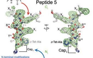
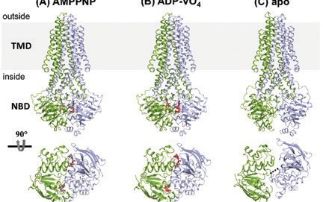
The Biological Cryo-Imaging Group was established a little over a year ago and brings together dedicated facilities for X-ray, light and electron microscopy at Diamond. The bending magnet B24 is the source of X-rays for the full field cryo-transmission X-ray microscope dedicated to biological X-ray imaging. The beamline has also established a cryo-super resolution fluorescence microscopy facility, which is a joint venture between Diamond and the University of Oxford. Exploiting electrons for imaging, the electron BioImaging Centre (eBIC) is the national centre for cryo-Electron Microscopy (cryo-EM) in the UK and provides a range of capabilities and supporting facilities for cryo-EM. The first year of the Biological Cryo-Imaging Group at Diamond has been a busy one, with both B24 and eBIC developing and expanding their capabilities. This has meant installation, commissioning of new instrumentation, and a major focus on recruitment. Read more  ...

  • annual review
    Unveiling RNA polymerase III transcription initiation with cryo-EM

    Unveiling RNA polymerase III transcription initiation with cryo-EM Jul 26, 2019

    RNA polymerase (Pol) III is a major determinant of lifespan in eukaryotes (organisms whose cells have a nucleus enclosed within membranes, unlike bacteria). The level of Pol III transcription is tightly linked to the rate of growth, as it is known to play a role in cancer, and neurodegenerative diseases. A better understanding of the structure of Pol III could lead to new therapies for these diseases. However, Pol III is the most complex nuclear RNA polymerase.

  • annual review
    Insight into work of molecular machines supporting genome stability

    Insight into work of molecular machines supporting genome stability Jul 23, 2019

    The work recently published in the journal Science by a team of scientists from Imperial College London featured the structure of the SWR1:nucleosome complex at 3.6 Å resolution, determined using cryo-Electron Microscopy (cryo-EM) data collected at the UK national electron Bio-Imaging Centre (eBIC).

  • Contact Us
  • About Diamond Light Source
  • Procurement
  • Supply Chain Transparency
  • Cookie Policy
  • Website Terms of Use
  • Privacy Notice

Diamond Light Source

Diamond Light Source is the UK's national synchrotron science facility, located at the Harwell Science and Innovation Campus in Oxfordshire.

Follow us on Facebook Follow us on Linkedin Follow us on Instagram Follow us on Bluesky

Diamond Light Source Ltd
Diamond House
Harwell Science & Innovation Campus
Didcot
Oxfordshire
OX11 0DE

See on Google Maps

Copyright © Diamond Light Source. Diamond Light Source® and the Diamond logo are registered trademarks of Diamond Light Source Ltd

Registered in England and Wales at Diamond House, Harwell Science and Innovation Campus, Didcot, Oxfordshire, OX11 0DE, United Kingdom. Company number: 4375679. VAT number: 287 461 957. Economic Operators Registration and Identification (EORI) number: GB287461957003.

feedback