Gianfelice Cinque, Village Coordinator
Soft condensed matter research at Diamond encompasses a range of complex organic molecules, biomedical specimens and inorganic systems. The Soft Condensed Matter Village can provide analysis at all scales of biology and medicine starting from individual proteins, to higher order structures such as fibers and whole cells. Access to the full range of length scales provides insights into fundamental life sciences, and tools for new molecule and drug discovery. In addition, the chemistry and physics of synthetic materials such as polymers, colloids, composite materials, foams, gels, organic semiconductors and liquid crystals are probed, and studies in areas such as catalysis, biomineralogy, thin films and surface phenomena are carried out within the village.
Studying the flexibility of the RNA-dependent RNA polymerase of influenza virus
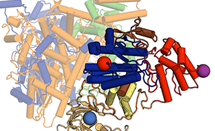 Influenza viruses are a major public health issue, with seasonal viruses responsible for approximately half a million deaths worldwide each year and new pandemic viruses constantly threatening to emerge. Although well-known pathogens, the way that influenza viruses replicate is not yet understood at a molecular level. The viruses copy their RNA genome using a viral RNA polymerase, therefore understanding the structure of this polymerase is fundamental to understanding the mechanism of viral replication. Read more...
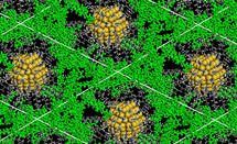
In situ single crystal synchrotron IR microspectroscopy of gas-solid reactions
 Highly porous materials such as metal-organic frameworks (MOFs) and zeolites are very effective at adsorbing gases and catalysing reactions, due to their ability to trap molecules in their pores. Infrared (IR) microspectroscopy experiments on the Multimode InfraRed Imaging and Microspectroscopy (MIRIAM) beamline, B22, have now revealed the processes involved in gas adsorption and catalysis in more detail than ever before. Read more...
Hybrid glasses: A new family of glasses captured with small angle X-ray scattering and calorimetry
 Hybrid glasses are a new variety of glass that have been discovered and reported in Nature Communications, assisted by in situ X-ray scattering experiments on the Small Angle Scattering and Diffraction beamline (I22). These new glasses comprise inorganic metals, like zinc, interlinked by organic bridges, such as immidazolate ligands. They have been obtained starting from metal organic frameworks (MOFs), an innovative range of novel micro-engineered crystalline materials. Read more...
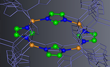
Making gold metamaterials with a twist
 Nanometer sized metal particles can have unusual optical properties, with benefits that include potentially ‘cloaking’ objects rendering them invisible. Ordered arrays of gold or silver nanoparticles that can interact with circularly polarised (CP) light – light where the electric and magnetic fields rotate rather than vibrate – would extend potential application even further. CP light is used in many everyday objects, such as compact disc players and many mobile phone display screens. Read more...
Using microfluidics on beamline B21
Often, scientists are interested in how their molecules change in response to the addition of another small molecule, particularly for biological samples, where small molecules could be potential drug candidates. Principal Beamline Scientist on B21, Dr Robert Rambo, and his team have been looking at how microfluidics could provide reliable and rapid screening of the effects of small molecules on proteins by Small Angle X-ray Scattering (SAXS). Microfluidics is the design of systems in which low volumes of fluids can be processed automatically for high-throughput screening. Fluids can be precisely moved, mixed, or separated and are controlled by their confinement in small, typically sub-millimeter channels where capillary forces play a role.
Work on this new tool started in 2015. A bespoke microfluidics cell was designed to enable users to move directly from the purification stage of producing their sample protein molecules, to mixing it with a small molecule inside a microfluidics cell. The cell has two micro channels, around 800 microns in size. One is connected by tubing to a high-performance liquid chromatography system and the other to an automated syringe, where a small molecule can be introduced to the custom cell. When this happens a laminar flow of the two streams is formed that will only interact at the point where they reach the X-ray beam, allowing for detection of data from the reaction point.
The team designed a stainless steel microfluidics cell to enable direct mixing of samples during the SAXS measurements. But they realised that a low-cost, disposable option was important for experiments where the cell would be difficult to reuse, such as where gels are formed in reactions or where trace metals are being analysed within living cells. The solution has been to use a 3D printer to produce unique sample cells for specific scientific investigations, which are printed on site using high-quality methyl acrylate polymers. While most B21 users can use the standard instrumentation configuration, the ability to make and print cells at the beamline is a useful additional tool.
The technique is in its infancy but so far a range of experiments have been undertaken. One example is the study of Amyloid fibrils by Professor David Middleton from Lancaster University and colleagues, which continues previous work they carried out at Diamond1. Amyloid fibrils are aggregates of misfolded proteins, whose formation has been implicated in Alzheimer’s and Parkinson’s diseases. Using a microfluidics experiment, the team hope to better understand the factors that promote or inhibit amyloid development in the presence of biological initiators.
For 2016, B21 will be offering experiments using microfluidic chambers as part of the beamline standard tool kit.
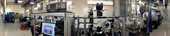
Figure 1: Panorama of the MIRIAM beamline, with the AFM-IR setup at the centre.
Coupling an AFM to an IR beamline gives the world’s first Synchrotron Photo-Thermal Nanospectroscopy
In 2015, the MIRIAM beamline (B22) demonstrated the world’s first example of Photo-thermal NanoSpectroscopy via Synchrotron IR. The technique combines three methods: synchrotron infrared (IR) microspectroscopy, atomic force microscopy (AFM) and photo-thermal detection, to create quantitative spectroscopic data at the nanoscale and beyond the diffraction limit of light2.
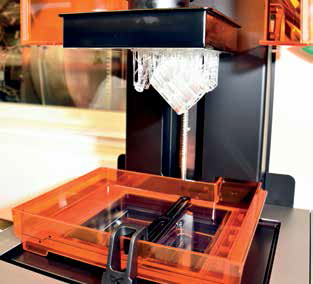
Figure 2: Rapid prototyping of sample environments using a laser focused, photopolymerization printer on B21.
IR spectroscopy is a non-destructive method that uses the IR vibrational excitation of chemical bonds to provide quantitative molecular information. It is used widely to study both organic and synthetic materials, spanning from the biological, chemical and physical sciences as well as multidisciplinary applications. Using the brighter and spectrally broader light emitted from a synchrotron source like Diamond, and coupling it into an IR microscope, has the advantage of opening up the microscopic scale; the spatial resolution and spectra quality are enhanced, whilst there is a dramatic increase in the speed of the experiments. Two of its exemplary uses are in biomedicine; to test cancer cells’ response to new therapeutic drugs, and following up stem cell differentiation by biochemical agents with regards to future regenerative medicine. But there is still the light diffraction limit. Optically microspectroscopy is unable to clearly resolve structures below the wavelength scale. For example, the optical method is unable to spatially discriminate the organelles inside a biological cell, or clearly image its “internal machinery”.
The new method developed by the team led by Dr Gianfelice Cinque is now overcoming this optical limitation using the close up near field detection offered with AFM. This technique uses a very fine tip on a oscillating cantilever arm to scan across the surface of a sample illuminated by the IR light, and give accurate molecular information of the microsample. It has a resolution of 100s of nanometers, orders of magnitude better than the optical light limit whilst maintaining the high specificity of IR spectroscopy to molecules.
The key to the Diamond team’s innovation is to use the AFM and photothermal nanoprobe detection to measure IR absorption and the subsequent mechanical expansion induced in the microsample. They shine the IR microbeam into the sample from below with the AFM mounted above in contact with the sample’s surface. Photons then absorbed by molecules in the sample will resonate, which causes thermal excitation. This means that there is a very local temperature rise (less than a hundredth of a degree), and thermal expansion (less than the size of an atom), which can be picked up by the AFM tip.
To amplify the signal, the team made use of resonance effects. In one of their biggest technical challenges, they added an ultra-fast mechanical chopper to the setup, designed to create short light pulses at the timescale needed to maintain the local excitation. When the chopper is tuned to work in resonance with the AFM cantilever, this produces a larger probe oscillation. The AFM signal can therefore be used to produce an absorption spectrum, giving an IR fingerprint of the molecular composition.
The team has tested this new photo-thermal IR method, which is quantitative and nanoscale sensitive. Initial experiments confirm it can give sub-wavelength spatial resolution at the 100 nm scale, above one order of magnitude better than optical IR microscopy. It can be applied to micrometer specimens, and is particularly useful for polymer and soft matter micro feature characterisation, including nanometric composite materials. The final target is to have a whole IR image of a microsample at nanoscale resolution, within a minute, including full molecular information.
The potential importance of this advanced spectroscopy technique is its ability to probe inside cells, something currently difficult to do. The resolution now possible should allow scientists to examine single cell interiors and the biochemical machinery and processes at the sub-cellular level. This includes the interactions and effects of viruses and bacterial infection and cell responses to new drug candidates. There will also be applications in materials science, probing nanoscale materials to examine, for example, how local physical morphology and chemical bonding alter with changing temperature or pressure. This novel nanospectroscopy and nanoimaging method is one of the main in-house research activities of the beamline staff.
I22 update
In 2015 a new L shaped detector was added to I22, for wide angle scattering (WAXS) data collection. The 2D nature of the WAXS data allows information on any anisotropic character in the scattering pattern to be observed. This gives ordering and orientation information that can be valuable when trying to understand rearrangements of structures, such as the behaviour of liquid crystals or polymers.
The inclusion of this new wide angle detector allows both wide angle and small angle scattering measurements, so molecular and atomic ordering data can be collected simultaneously. This capability will provide added value to users’ experiments.
In the first half of 2016, a software update will allow users near to real-time access to reduced data. This improvement will allow intelligent experimentation – the ability to modify studies during experiments, depending on results.
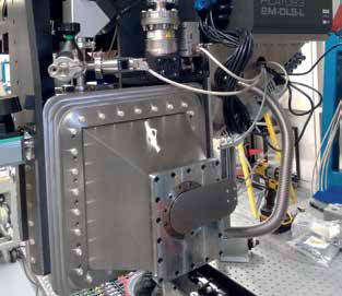
Figure 3: The new detector on I22.
B23 update
B23 is used extensively to characterise the structure in solution of biologically important molecules and their interactions with other molecules. This is particularly useful for protein structures that are natively disordered and cannot be crystallised or where nuclear magnetic resonance (NMR) spectroscopy does not produce good results, as is often the case for membrane proteins and large protein complexes. It can enhance the understanding of the molecular mechanisms of diseases and can contribute towards the development of rational therapeutic strategies. It is also useful for screening proteins binding with other proteins (protein-protein interactions) and with drug-like molecules, which is crucial in identifying potential new therapeutic targets.
B23 is the only beamline in the world to offer circular dichroism (CD) imaging capabilities that cannot be achieved using bench-top CD instruments. Staff scientists work closely in partnership with users to optimise the set-up parameters and produce the best results possible. B23 is constantly upgraded to improve the sample chamber unit and in-house software for data processing and analysis to enhance the user experience.
In 2015 the unique capability to use 96- and 384-well plates for high throughput CD (HTCD) was successfully established. For 2016, a new sample chamber is planned, including a camera to couple spectroscopy with microscopy to further enhance the CD imaging capability of B23. Preliminary results of CD imaging have been used for time resolved SRCD studies, developed to understand the early stages of protein misfolding responsible for neurodegenerative conditions such as Alzheimer’s and Parkinson’s diseases. Understanding the mechanism is the key step towards the development of rational therapeutic strategies.
References:
- Madine J., Davies H. A., Hughes E. and Middleton, D. A. Heparin Promotes the Rapid Fibrillization of a Peptide with Low Intrinsic Amyloidogenicity. Biochemistry 52, 8984-8992 (2013).
- Donaldson P.M., Kelley C.S. , Frogley M.D., Filik J., Wehbe K. and Cinque G. Broadband near-field infrared spectromicroscopy using photothermal probes and synchrotron radiation. Optics Express 24, 3, 1852-1864 (2016).
The village’s four beamlines serve differing, although sometimes overlapping, user communities, from both the life and physical sciences, as well as fields such as archaeology, cultural heritage, environmental and forensic science. Users often share an interest in complex systems, and studies are focused on how three dimensional molecular structures undergo compositional changes in dynamic conditions, providing crucial information on how structure and composition is linked to function.
The village includes the diffraction beamlines I22 and B21, which both make use of Small Angle X-ray Scattering (SAXS), with B21 focusing on solution-state scattering, and I22 providing structural investigations into a wide range of materials. Studying the elastic scattering of low wavelength (0.06-0.35 nm) X-rays, recorded at very small angles, provides information on shape, size and ordering of materials at the nano level, without the need for the atomic ordering found in crystals.
Beamline B22 provides Multimode Infrared Imaging and Microspectroscopy (MIRIAM), which gives quantitative analysis of the specific molecular composition and microscopic spatial distribution over a large spectral region in scanning microscopy or imaging mode, including far-infrared (or TeraHertz) spectroscopy, as uniquely exploited at Diamond. Beamline B23 studies circular dichroism (CD) in complex systems – a spectroscopic technique for probing chiral materials that exist in different conformations and, particularly for proteins, how this affects their function.
The Highly Automated Throughput SAXS beamline, B21 (HATSAXS), is dedicated to the study of particles in solution, with a majority of the users coming from the biological sciences community. SAXS is an alternative to diffraction techniques, where crystallography is not possible, and provides a structural measurement of the thermodynamic state of the sample. It is an ideal technique for biomolecules in solution as these molecules can be tested under a wide range of experimental conditions. SAXS has also proven useful for the study of synthetic materials such as quantum dots (particles used in active display technologies). SAXS is particularly sensitive to changes in the sample structure and can provide unique insights into the behaviour of biological molecules that can only be observed in solution, including large conformational changes and transient protein-protein interactions
At the start of 2017, B21 will be upgrading its X-ray optics with an expected eighty-fold increase in X-ray intensity. Coupled with the exceptionally low background scatter, the increased intensity will push the sample concentration requirements substantially lower and enable sub-second measurements.
The Small Angle Scattering beamline I22 (SAPPHIRES) provides combined small and wide angle scattering studies (SAXS and WAXS) on a wide variety of low order biological and synthetic molecular assemblies. It is useful for gaining information on partially ordered materials, giving information on pore sizes or lamellar, fibrous or helical structures. As well as biological and physical studies, SAXS is also proving important in environmental and conservation sciences.
Experiments can probe structural changes in a variety of changing sample environments – for example, raising and lowering the temperature, increasing the pressure and stretching samples, changing gas flow and changing magnetic and electric field effects. Results can also be obtained in millisecond timescales to explore kinetic processes such as protein folding events, nucleation and growth of crystals and structural evolution in polymers and colloids.
The MIRIAM beamline (B22) provides Infrared (IR) Microspectroscopy, which allows sensitive quantitative molecular identification and spatial resolution at the highest resolution optically attainable. The technique has applications in many fields including in situ analysis of catalysts, advanced inorganic-organic combined materials, polymers under stress-strain, pharmaceutical drugs testing on cell cultures, histopathological tissue section analysis, as well as analysis of ancient artefacts.
The IR radiation available at the MIRIAM beamline spans from the near-IR up to the far-IR (or THz) region. For users, this allows the finest identification of functional groups and biochemical characterisation via IR maps in two dimensional samples at the micron scale in subminute experiments, to the study of ultra-low energy excitations and conformational changes in large molecular complexes with far IR photons. The IR microprobe can pinpoint single cancer cells’ response to chemotherapy much earlier than any visible microscopy method used in biomedicine, or highlight environmental changes across the microlayers of ancient/modern painting fragments, or study new superconductor’s pseudo energy-gap, or structural changes in engineered microcatalysts in operando.
Over the last few years, the optimisation of far-IR spectroscopy at Diamond has expanded with experimental capabilities specifically in the ‘THz gap’ domain - the region of the electromagnetic spectrum between radiowaves and IR light where there are few technologies for generating/detecting radiation. For the sub-TeraHertz domain, IR coherent light is produced by very short electron bunches circulating in Diamond operating in the low alpha special beam mode: this is regularly available for the broadest THz research community, and has been successfully exploited for disentangling metalorganic- framework molecules dynamics, also in combination with molecular simulations and neutron experiments at ISIS.
B22 is progressing with the optimisation of the Focal Plane Array (FPA) detector, already available for fast IR imaging. The beamline plans to develop dedicated optics to expand the field of view and adapt the IR Synchrotron beam to operate at the different magnifications available at the IR microscope, namely: from 15 times and 20 times magnification, suitable for 100 μm scale extended samples, up to 36 times and 74 times magnification, suitable for submicron imaging via oversampling.
Running since 2009, the beamline for Synchrotron Radiation Circular Dichroism (SRCD), B23, allows users to characterise the structure of a wide variety of materials in solution and in solid state films, using circularly polarised light. Molecules that are chiral, that is they have two mirror-image versions, will absorb circularly polarised light differently. This property, known as circular dichroism (CD), can be used to very quickly characterise the conformation of chiral polymers and biopolymers such as nucleic acids and carbohydrates, as well as the folding of proteins. As conformational structure can also dictate function and properties, CD is an important technique in biomedicine and materials science.
For materials science, the unique highly collimated small light beam of B23, 0.5 to 1 mm diameter, is used for spatially resolved studies enabling the SRCD mapping of material films, a crucial step in the development of new materials. The method has commercial applications in allowing materials' properties to be optimised in terms of sample preparation and production.
Diamond Light Source is the UK's national synchrotron science facility, located at the Harwell Science and Innovation Campus in Oxfordshire.
Diamond Light Source Ltd
Diamond House
Harwell Science & Innovation Campus
Didcot
Oxfordshire
OX11 0DE
Copyright © Diamond Light Source. Diamond Light Source® and the Diamond logo are registered trademarks of Diamond Light Source Ltd
Registered in England and Wales at Diamond House, Harwell Science and Innovation Campus, Didcot, Oxfordshire, OX11 0DE, United Kingdom. Company number: 4375679. VAT number: 287 461 957. Economic Operators Registration and Identification (EORI) number: GB287461957003.