Kawal Sawhney, Village Coordinator
Synchrotron radiation has revolutionised our understanding of materials through X-ray diffraction and imaging techniques. The Materials Village at Diamond continues to provide the tools to extend and broaden our current knowledge, from probing materials designed for energy storage and carbon capture to imaging natural biological and geological structures.
Faster, higher resolution phase-based 3D X-ray images with a beam tracking approach
 Conventional X-ray imaging is based on the attenuation of X-rays, a reduction in intensity as the beam passes through matter. Some features are considered to be ‘X-ray invisible’, due to the limited attenuation they cause. Recent research has focused on phase effects, which can enhance detection of these features, and also enhance the visibility of the image in general. ‘Dark field’ or ‘ultra-small angle scattering’ is an additional phase-related X-ray mechanism under investigation, which can provide complementary information on the sample being studied. Read More
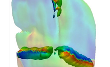
Quantifying bone bridging across longitudinal growth zones in the tibia during healthy and pathological ageing
 Osteoarthritis (OA) is a debilitating disease and worldwide healthcare burden. It is characterised by the loss of articular cartilage that normally covers the ends of bones to allow pain free movement. Current clinical strategies only manage the joint pain and do not address the underlying molecular mechanisms that trigger and fuel this degenerative disease. Prior studies have suggested that cells in the articular cartilage undergo uncharacteristic changes, which may result in the deterioration of this tissue. However, little is known of the involvement of a further type of cartilage critical for bone lengthening, known as growth plate cartilage. Read More
New imaging technique to map multiple elements during the solidification of alloys
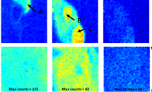 A new imaging technique was designed and tested that maps the distribution of multiple elements during a dynamic process. The dynamic process explored here is the solidification of a metal alloy, but the technique has applications throughout metallurgy and materials science. An aluminium alloy containing silver (Ag), zirconium (Zr), and molybdenum (Mo) was melted at 700 °C, before re-solidifying. As the metal freezes, metallic crystals (grains) grow. During this freezing, the alloying elements and impurities segregate and become concentrated in the remaining, rapidly vanishing liquid. In these last-to-freeze regions, highly concentrated clusters of elements freeze into different types of tiny crystals. These give the metal an irregular microstructure which can have strong negative effects on the alloy mechanical properties and recyclability. This frozen microstructure is hard to modify, making it commercially important to understand and manipulate the way elements distribute and segregate during solidification. Read More
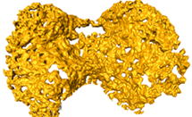
How do dislocations shuffle to grow crystals?
 Bragg Coherent Diffraction Imaging (BCDI) is a technique which became available for X-rays with the introduction of 3rd generation, undulator-based storage rings like that of Diamond Light Source. The BCDI method images crystals in three dimensions (3D) by solving the phase problem using iterative algorithms, and then inverting, the diffraction pattern surrounding one of the Bragg peaks diffracted by the crystal under examination. The particular advantage of BCDI is phase contrast imaging, where the phase of the 3D image of a crystal is attributed to the local distortions of the crystal due to the presence of lattice strains. The work of Johannes Ihli, Jesse Clark and colleagues, published in Nature Materials and reported here, used BCDI to observe screw dislocations by their characteristic spiral-shaped distortion seen within growing crystals of calcite. The dislocations were seen to move ahead of the growing {104} facets of a calcite crystal, grown in situ from solution at the beamline. Read More
Controlling the warp and weft of molecules that are knitted together
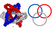 The Small Molecule Single Crystal Diffraction beamline (I19) has helped elucidate a novel interlocked molecular structure made up of three interlinked rings, like a nanoscale version of chainmail. Such interlocked molecular structures could lead to materials with novel properties, but they have proved difficult to produce because of a paucity of methods for weaving or knotting multiple chains of atoms together so they can’t unravel or unlink. Producing the new interlinked ring structure, termed a cyclic [3]catenane, thus required the development of a completely new synthesis method. Read More
Small Molecule Diffraction beamline (I19) upgrade
2015 saw the upgrade of the I19 beamline chemical crystallography diffractometer, which has been in operation since 2008. The upgrade was designed to improve beamline capabilities and allow users to collect data from smaller, more weakly diffracting crystals than was previously possible. At the same time, the aim was to create more efficient and faster data collection.
The new instrument was designed by a small team from Diamond’s Mechanical, Electrical, Motion and Controls Groups with input from the I19 beamline scientists. The fabrication of the new diffractometer involved a number of local companies and its overall design was based on experience with the operation of the old instrument. The design also borrowed heavily from the sample viewing and beam conditioning equipment that was also developed at Diamond for the macromolecular crystallography (MX) beamlines.
The new diffractometer has the conventional three-circle geometry and incorporates a unique dual air-bearing system. An air-bearing, which is composed of a shaft floating on a film of high-pressure air within a cylinder, has superior accuracy and stability over a conventional mechanical bearing and this allows the sample to be more accurately positioned. Together with a tightly focused beam and improved collimation, this is allowing data to be collected from very small crystals, in the 5 micron range.
The increased efficiency of data collection, and the ability to collect data faster, has been achieved by incorporating a photon-counting Pilatus 2M detector to replace the CCD (charge coupled device) detector used on the old instrument. The Pilatus detector works in a ‘shutterless’ mode that allows the diffraction data, in the form of 2D images, to be read continuously, while the diffractometer scans (rotates) the sample at a constant speed. This is much more efficient than the CCD detector where each image was taken discretely, requiring an X-ray shutter to accurately time the exposure, and with the diffractometer performing discrete steps for each image in the scan. The time overhead between each image was appreciable, often longer than the exposure for each image.
The new diffractometer can now perform a data collection in approximately five minutes rather than the 90 minutes required previously. This will not only increase the sample throughput, but will also offer users the opportunity to perform parametric experiments in much smaller intervals, such as studying the variation of a crystal structure with temperature.
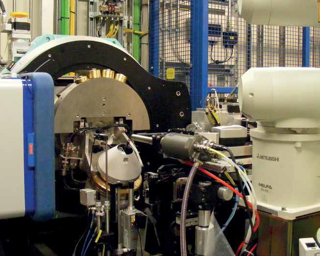
Figure 1: Upgraded Experimental Hutch 1 on I19.
In addition, the new detector’s larger area allows it to be positioned further from the sample than was previously possible with the CCD detector, while maintaining the angular range of the diffracted data. This reduces the recorded background scatter considerably and allows very weak diffracted intensities to be discerned above a much lower background.
The upgraded diffractometer became available to users in January 2016 and, when the robotic sample changer is recommissioned, it will be possible for users to operate the beamline remotely from their home institution.
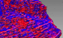
X-ray speckle-based technique – a new way to explore coherence properties
The flexibility of the B16 Test beamline allows Diamond scientists to constantly improve and innovate. An example of this is the development of an X-ray speckle based technique using the near-field speckle phenomenon, which is widely exploited in laser interferometry. In a recent study, the coherence properties of X-ray beams have been measured by analysing the spectrum of X-ray near-field speckle patterns1. In addition, simply by using a piece of sandpaper, the Diamond team have devised a new imaging technique that could eventually lead to cheaper and safer X-ray imaging for medical applications.
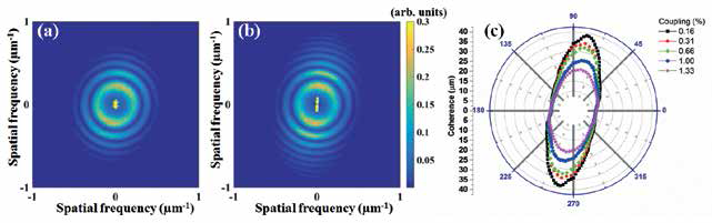
Figure 2: Power spectrum of the speckle in (a) 1.33% and (b) 0.16% coupling conditions. (c) two-dimensional transverse coherence distribution for the vertical electron-beam coupling (C) at 0.16%, 0.31%, 0.66%, 1.00% and 1.33%.
Speckle patterns are the product of wavefront interference, produced by the diffuse reflection of a highly coherent light source by a rough surface or inhomogeneous object. The pattern generated appears to be a randomintensity distribution of light that gives the surface a granular appearance. Initially, the technique was developed to test the quality and performance of the X-ray optics employed on the Diamond beamlines, including their coherence – that is, the phase uniformity of the X-ray wavefront2. This property is not easy to measure, but this new technique provides a sensitive and simple method.
Being able to measure the coherence of Diamond’s X-rays is important to understanding the characteristic optics of each beamline and is also useful for many advanced experimental techniques such as Coherent X-ray Diffraction, X-ray Phase Contrast Imaging and X-ray Photon Correlation Spectroscopy. But so far it has not been simple to do, as it required high-precision optical elements or multiple measurements. That is until Diamond scientists, led by Dr Kawal Sawhney, looked at exploiting speckle images. Placing a porous membrane (with 0.8μm pores) in front of the detector, speckle images could be taken and from these, the coherence of the X-ray beam quantitatively calculated. As shown in Fig. 2, the method was validated for a range of X-ray source sizes produced from Diamond’s electron beam.
The success of the method leads the Diamond team to consider how speckle images could be used for creating X-ray 2D phase contrast images3,4,5. Their method required one further tool – a piece of textured sandpaper. The speckle pattern is firstly generated with the piece of sandpaper in front of the beam, and recorded by the detector. Then a sample is placed in front of the sandpaper, and the process repeated to give a second series of speckle images. Analysis of the changes in the speckle images using a digital image correlation algorithm allowed an image to be built up. The technique is able to pick out differences between materials with very similar X-ray absorption, with better image resolution. The method is particularly useful for imaging contrasts in biological soft tissue which are difficult to differentiate with conventional techniques.
The initial work carried out on B16 has been further developed to create 3D tomographical imaging capabilities. In 2015, Diamond scientists worked together with a team from the PETRA III Synchrotron in Hamburg to produce the first 3D speckle-based images of a human artery5. The images produced had enhanced contrast compared to conventional X-ray absorption imaging.
X-ray near-field speckle imaging promises better contrast for biological imaging of soft tissue at lower X-ray dosages. This, and its simplicity and speed of measurement, make it attractive for conventional medical X-ray imaging. The team are continuing to develop the technique on the B16 beamline and the method could ultimately be developed as a table top device.
References:
- Kashyap Y., Wang H. and Sawhney K. Two-dimensional transverse coherence measurement of hard-X-ray beams using near-field speckle. Phys Rev A 92, 3. (2015).
- Wang H., Sutter J. and Sawhney K. Advanced in situ metrology for X-ray beam shaping with super precision. Opt Express 23, 2, 1605-1614 (2015).
- Wang H., Kashyap Y. and Sawhney K. Hard-X-Ray Directional Dark-Field Imaging Using the Speckle Scanning Technique. Phys Rev Lett 114, 10, 103901 (2015).
- Wang H., Berujon S., Herzen J., Atwood R., Laundy D., Hipp A., et al. X-ray phase contrast tomography by tracking near field speckle. Sci Rep 5,8762 (2015).
- Wang H., Kashyap Y. and Sawhney K. From synchrotron radiation to lab source: advanced speckle-based X-ray imaging using abrasive paper. Scientific Reports 6, 20476 (2016).
The work supported by the Materials Village continues to provide a greater understanding of the all-important relationship between structure and function. This underpins the chemistry and materials science of strategically important areas such as pharmaceutical science, supramolecular chemistry, magnetic and biological materials. Diamond offers beamlines with highly coherent X-rays that use phase effects to image structures that are not visible in normal radiography and that cannot be easily imaged in a laboratory setting.
The Materials Village brings together four beamlines that offer a wide range of techniques for studying materials. The Small Molecule Diffraction beamline (I19) supports X-ray crystallography using single-crystal methods for studying molecular structures. The Materials and Magnetism beamline (I16) provides a unique, world-class single crystal X-ray diffraction facility for probing magnetic properties, and the X-ray Imaging and Coherence beamline (I13) facilitates non-destructive imaging of materials and their internal features at the micro and nano scale. The village also includes the Test beamline (B16), which is used to test new equipment and instrumentation, and develop new experimental methods.
Single-crystal X-ray diffraction is the definitive way to determine a material’s molecular structure. I19 can provide optimised results for small crystals and complex molecular structures that are weakly scattering, and where results are difficult to obtain from other X-ray sources. The beamline also allows structural changes to be studied under a variety of environments such as variable pressure or temperature, under photo-excitation or gas exchange. The beamline supports the study of electronic ordering and the structure of excited systems.
2015 saw a significant upgrade in I19’s capabilities, with the installation of a new end station, including a new detector. This became available to users in January 2016 and the beamline now has the means to analyse crystals as small as 5 microns, with data collection almost 20 times faster. In the coming year, the integration of software for the robotic control of samples will support remote use of the beamline.
The Materials and Magnetism beamline provides a unique, single crystal X-ray diffraction facility for high resolution diffraction and scattering experiments focused on the measurement of magnetic and electronic ordering effects. The beamline is optimised for measurements on very small sample volumes, including artificial nanostructures, thin films and surfaces. The beam can be focused to a spot less than 50 by 200 microns, with a continuously tunable X-ray energy. Samples can be analysed at temperatures from as low as 6 K to over 600 K, giving access to a wide range of electronic phases. Very low (down to 2 K) temperatures can be achieved, in horizontal scattering geometry, with the beamline’s pulsed-tube cooler.
2015 developments on I16 include replacement of the diffractometer control system with a new Diamond- standard solution that can be maintained and enhanced in-house, and provides highly efficient and accurate continuous scan modes as well as improved control. Software developments have also enhanced beamline capabilities and have allowed the beamline to write full data files in Nexus, the Diamond standard data format, which facilitates 3D diffraction data mapping, for thin films studies, diffuse scattering and heterogeneous structures.
At 250 metres in length, I13 is Diamond’s longest beamline and the one village facility dedicated to non-destructive imaging. The long length allows for more vertically and horizontally coherent X-rays, meaning their wave phases stay constant – an important property for a number of techniques. The beamline’s ability to reveal the internal features of materials at the micro and nano length scale is being utilised by a broad range of scientists, including biologists, materials scientists, geologists, archaeologists and engineers. The beamline runs two simultaneous and complementary X-ray imaging branchlines – Imaging and Coherence. The coherence line uses Coherent Diffraction Imaging (CDI) methods. This uses laser-like pulses of coherent X-rays, whose diffraction patterns can be reconstructed into high resolution images.
I13 is partly operated in collaboration with the University of Manchester. The collaboration has been running since 2010 and gives the university speedy and streamlined access to Diamond’s imaging and tomography capabilities. Scans capture 3D images using partially coherent radiation which enables contrast enhancement at the edges of structures. This ‘in-line phase contrast imaging’ is particularly useful for biomedical applications where the contrast between structures is weak. Imaging of soft tissue is now possible and 2015 saw a remarkable study of a mouse knee, with the bone, cartilage and other soft tissue distinguishable. The study, by Professor Andrew Pitsillides of the Royal Veterinary College, University of London, looked at the knee joints of mice, genetically engineered osteoarthritis1. Using microtomography they were able to quantify the abnormal bone growth in the joints.
In 2015, a full-field X-ray microscope was tested, to improve spatial resolution to up to 50nm. This nano-imaging technique will be operational in the near future on the imaging branchline. The project was carried out in conjunction with Dr Christian David and Dr Jeroen Bosgrathe at the Paul Scherrer Institute in Switzerland, a world-leading facility for X-ray optics. In 2016, it is planned to upgrade the equipment on the coherence branchline, with the installation of robot arms to hold the detector, and an upgraded photon-counting EXCALIBUR detector. The upgrades will allow coherent diffraction experiments to be easier to run for the non-expert user community.
The final beamline in the village is Diamond’s Test beamline (B16). It is used to test new developments in optics and new detector technology without interrupting the schedule of other beamlines. The highly flexible nature of the beamline is designed to allow rapid responses to requests from visiting scientists, allowing them to develop proof-of-principle experiments and novel techniques. Working closely with Diamond’s Optics and Metrology Group, the beamline has had another successful year. Beamline upgrades in 2015 include a new high stability granite table, installed with associated rotation and translation stages which allow imaging and micro-diffraction studies. An optical microscope has also provided quick sample alignment and real-time optical imaging of samples.
The beamline is designed to develop proof-of-principle experiments for new techniques and 2015 saw the piloting of a new imaging method, which is featured in this report, to map the distribution of multiple elements during the dynamic solidification of alloys.
References:
- Staines K.A., Madi K., Mirczuk S. M., Parker S., Burleigh A., Poulet B., Hopkinson M., Bodey A. J., Fowkes R. C., Farquharson C., Lee P. D. and Pitsillides A. A. Endochondral growth defect and deployment of transient chondrocyte behaviours underlie osteoarthritis onset in a natural murine model. Arthritis & Rheumatology (2015).

Diamond Light Source is the UK's national synchrotron science facility, located at the Harwell Science and Innovation Campus in Oxfordshire.
Diamond Light Source Ltd
Diamond House
Harwell Science & Innovation Campus
Didcot
Oxfordshire
OX11 0DE
Copyright © Diamond Light Source. Diamond Light Source® and the Diamond logo are registered trademarks of Diamond Light Source Ltd
Registered in England and Wales at Diamond House, Harwell Science and Innovation Campus, Didcot, Oxfordshire, OX11 0DE, United Kingdom. Company number: 4375679. VAT number: 287 461 957. Economic Operators Registration and Identification (EORI) number: GB287461957003.