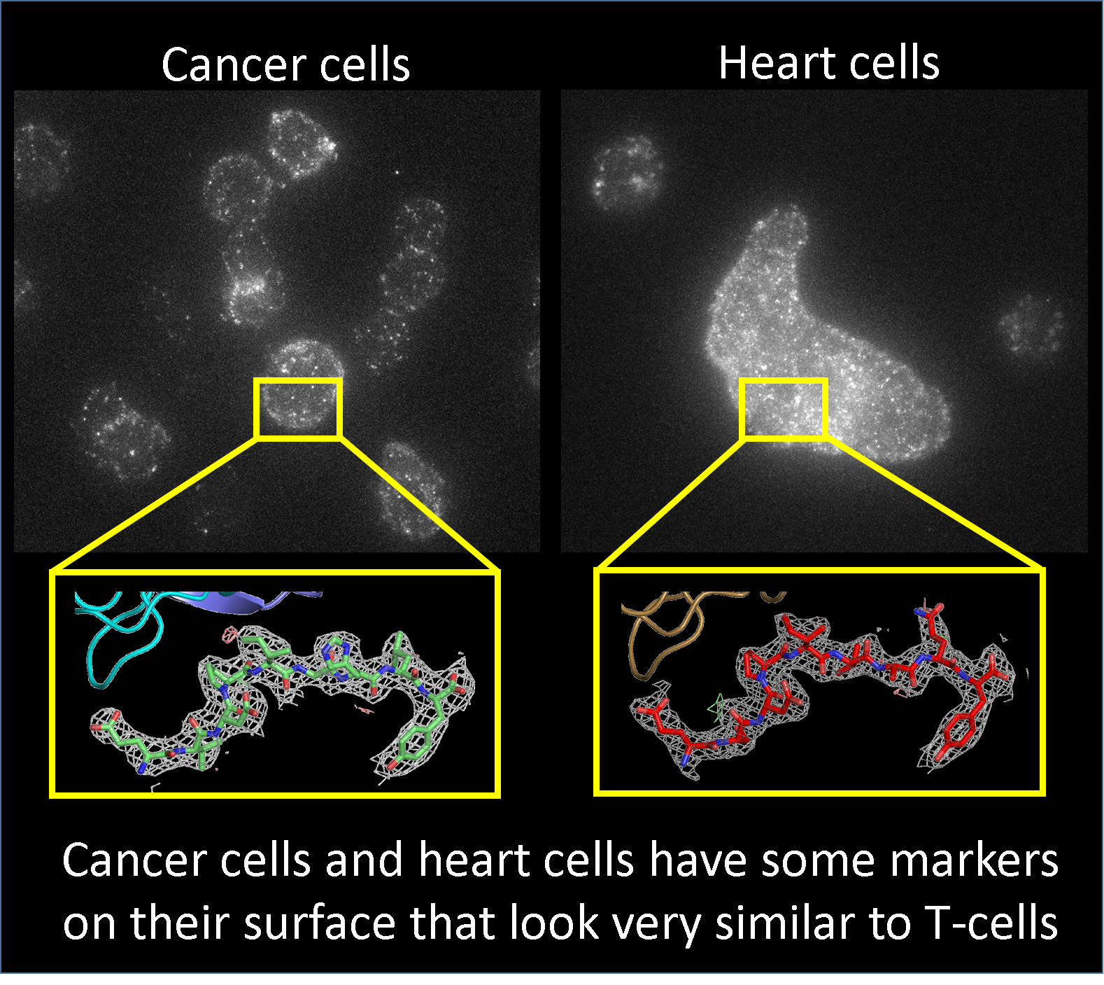
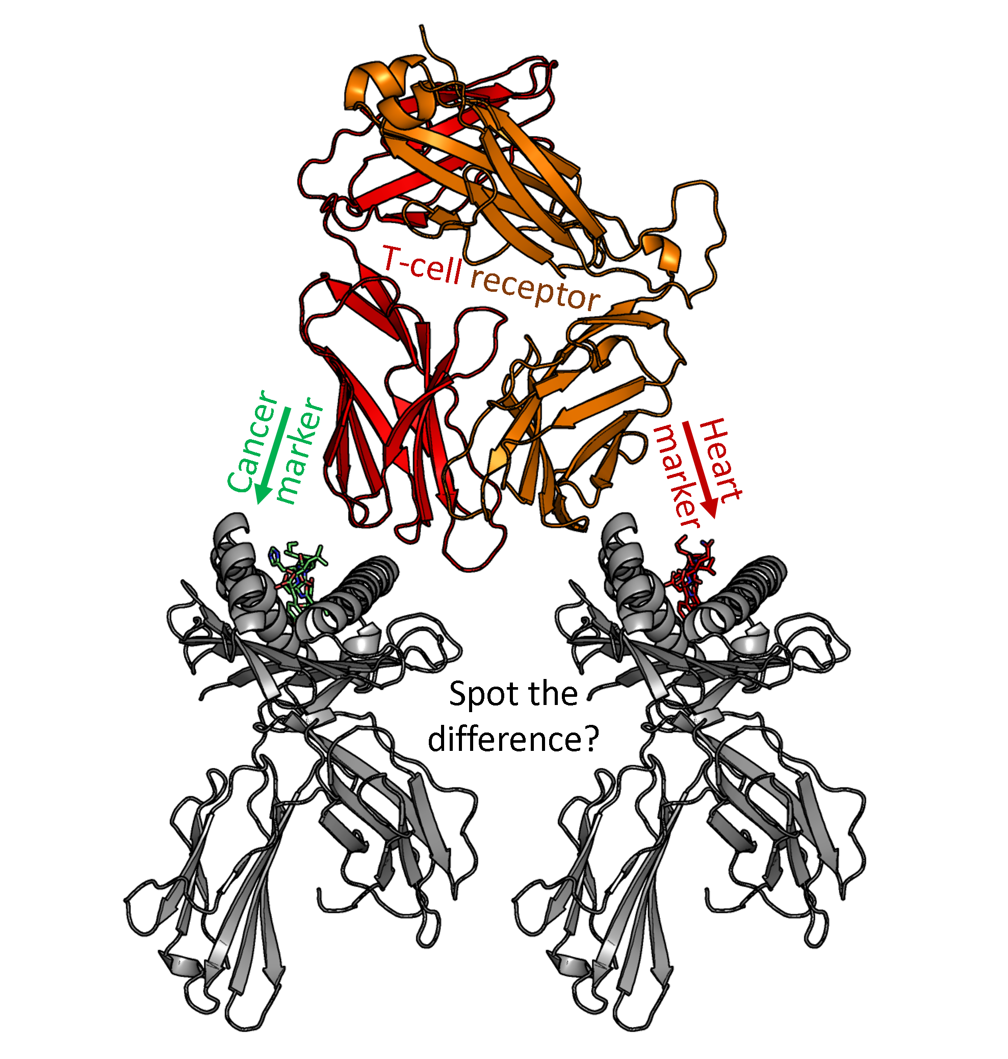
The synchrotron enabled the team to visualize this interaction between the engineered TCR and the cancer and heart tissue markers, to reveal that at an atomic level they were both similar in shape, making it extremely difficult for the T-cells to differentiate between the two.
Dr David Cole, from Cardiff University’s School of Medicine, senior author on the study, said: “This discovery is significant in a number of ways. Firstly, the images gleaned by the X-ray crystallography enabled us to directly reengineer the modified TCRs to significantly reduce its contact with healthy tissue, which is proof of concept for a safer, more effective design for a next generation of cancer drugs.
“Secondly, it shows how T-cells might cause unwanted damage to healthy tissue in other diseases such as type 1 diabetes, multiple sclerosis and rheumatoid arthritis. Moreover, the data explains, at the molecular level, why two patients suffered from cardiovasculardamage after receiving a novel cancer treatment – and how to avoid this from happening in future.”
Dr Pierre Rizkallah, lead author from Cardiff University’s School of Medicine, said: “The key to the new findings is the ability to visualise, at the atomic level, how the TCR ‘sees’ different markers, whether expressed on cancer cells or healthy cells. This is drug design on the scale of a protein, and X-ray diffraction is truly an incomparable tool in our hands for achieving these results.”
Professor Brian Baker, from the University of Notre Dame, said: “Modified T-cells are currently generating a huge amount of interest as a new breakthrough therapy to fight cancer. However, there is still much to learn about the potential side effects that these modified cells may have.
“The striking new study by Dr Cole and colleagues represents a very significant step in demonstrating why unanticipated side effects can occur, and how they might be avoided in future work, improving both safety and efficacy in cancer immunotherapy.”
The research was funded by The Wellcome Trust, with support from the facilities and staff at Diamond Light Source.
Atomic resolution cartoon picture of a modified T-cell receptor (red and orange) attempting to spot the difference between a protein marker found on the surface of some cancer cells (green and grey) and a similar shaped protein marker expressed on the surface of heart cells (red and grey).