From Dorothy to Diamond: Hodgkin’s Legacy
On 12 May 1910 Dorothy Hodgkin was born as Dorothy Mary Crowfoot in Cairo, eldest daughter of John Winter Crowfoot and Grace Mary Hood. During a career spanning seven decades she pioneered the technique of protein crystallography and applied X-ray crystallography to the study of small molecules of biomedical interest advancing our understanding of the chemistry of biological processes and antibiotic action. Today protein crystallography is applied at synchrotrons around the world, and Diamond has five beamlines dedicated to the technique. One hundred years after Hodgkin was born the Protein Database lists over 56,000 structures solved using the technique she pioneered.
![]() See A Modern X-ray Diffraction Experiment: Vitamin B12 Revisited
See A Modern X-ray Diffraction Experiment: Vitamin B12 Revisited
When Dorothy was two years old, Max von Laue was Professor of Physics at the University of Munich. Von Laue had been pondering the passage of light through periodic arrangements of particles. It occurred to him that X-rays, discovered by Wilhelm Röntgen 17 years previously, were supposed to be a form of electromagnetic rays with a shorter wavelength than visible light, and that passing these through a crystal would cause them to interact with the crystal in such a way that it would generate a pattern that depended on the crystal structure. Von Laue successfully recorded the first X-ray diffraction pattern, from a crystal of copper sulphate, worked out the mathematical formulation and published his work in 1912, which led on to his winning the Nobel Prize for Physics in 1914.
The nature of things
 |
| Cover of "Concerning the Nature of Things" |
Shortly after von Laue’s data was published, it caught the attention of William Lawrence Bragg, then working in Cambridge. After discussions with his father, William Henry Bragg, he set about re-interpreting the X-ray pictures published by von Laue’s group. He realised that a focussing effect seen by the German group would arise if the X-rays were reflected by planes of atoms in the crystal. This enabled him to reformulate von Laue's conditions for diffraction into what became known as Bragg's Law, which gives a direct relationship between the crystal structure and its diffraction pattern.
This made it possible to use X-ray diffraction to determine crystal structures, and the Braggs used their new technique to ‘solve’ the structure of a number of crystals, including sodium chloride, Calcite and Pyrites: X-ray Crystallography was born.
The Bragg father and son received the Nobel Prize for Physics in 1915, "for their services in the analysis of crystal structure by means of X-rays" when William Lawrence Bragg as just 25. In 1923 William Henry Bragg invited to present the Royal Institution Christmas Lecture. His topic was “Concerning the Nature of Things”, and in 1925 was published as a book for children. This was given to the 15 year old Dorothy Hodgkin, and sparked her lifelong fascination with crystallography.
“Broadly speaking, the discovery of X-rays has increased the keenness of our vision over ten thousand times and we can now 'see' the individual atoms and mo1ecules."Sir William Henry Bragg in "Concerning the Nature of Things"
In 1928 Dorothy started studying chemistry at Somerville College, Oxford. She attended a few lectures in X-ray crystallography, then in the Mineralogy Department, until the lecturer persuaded her to delay for a year whilst she improved her chemistry knowledge. Sadly the lecturer died and she was unable to continue formal training, but she did get to see a range of luminaries including Ernest Rutherford, Niels Bohr and Peter Debye, as well as a lecture by J D Bernal on the metallic state which contained a significant component of X-ray crystallography. After she graduated, Dorothy headed for Cambridge to work with Bernal.
It was in Cambridge in 1934 that Dorothy began her ventures into protein crystallography, following Bernal's lead in studying pepsin molecules. She used X-ray diffraction to establish that "the arrangement of atoms inside the protein molecule is also of a perfectly definite kind." Interestingly, the same experiment was the first to show that exposure to X-ray radiation damages the crystal - a problem crystallographers still face today.
The birth of structural biology
Not long after these results were published Dorothy returned to Oxford. In 1935 she grew crystals of insulin, and was delighted when on developing the X-ray photograph, she saw a regular array of tiny spots, indicating underlying atomic order within the crystal. However, it would be another 34 years before the tertiary structure could be solved.
"I used to say the evening that I developed the first x-ray photograph I took of insulin in 1935 was the most exciting moment of my life. But the Saturday afternoon in late July 1969, when we realized that the insulin electron density map was interpretable, runs that moment very close."Dorothy Hodgkin
Insulin was a particularly complex molecule at that time, and in the intervening years, alongside teaching and bringing up a family, Dorothy went on to solve the 3D atomic structures of a string of other important biological molecules; starting with cholesterol in 1937, penicillin eight years later and vitamin B12 in 1954. She received the Nobel Prize in Chemistry in 1964 for her work on penicillin, but this also recognised that her research had helped pioneer a whole new field - structural biology.
As structural biologists were realising the power of using X-ray diffraction to determine protein structures, the field of particle physics was also rapidly advancing. The end of the Second World War was something of a golden era, as both scientists and resources were released from the war effort. Prior to the war, cyclotrons were at the cutting edge of particle accelerators, but as physicists demanded higher and higher energies, the effects of relativity became significant. Cyclotrons were not able to support relativistic particles, and in 1945 the physicist Edwin McMillan proposed a new machine, the electron synchrotron, which would allow their relativistic acceleration.
The first of the machines to operate was in Woolwich, UK in 1947, quickly followed by a machine built by General Electric (GE) in Schenectady in the US. In the GE model, the electrons travelled in a glass vacuum chamber and so it was here that a new phenomenon was observed for the first time: synchrotron light. The existence of synchrotron light had already been predicted, but the conditions that would determine the frequency were unknown, so it was sheer coincidence that this machine generated visible light.
As synchrotrons increased in size to accommodate higher energy particles, the dominant frequency of radiation shifted into the X-ray region. However, synchrotron radiation continued to be seen as a waste of energy, to be eradicated as far as possible. In 1956 Tomboulian and Hartman were granted two weeks’ use of a synchrotron at Cornell, where they not only confirmed the spectral and angular distribution of synchrotron light, but also carried out an X-ray spectroscopy experiment parasitically attached to the side of the machine.
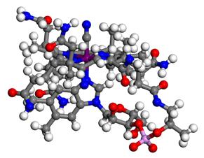 |
| The structure of vitamin B12 |
By this time, as well as Dorothy's work determining the structure of penicillin and vitamin B12, Franklin and Wilkins used fibre X-ray diffraction to record measurements for DNA, measurements that were incorporated into the 3D model of DNA by Watson and Crick. Meanwhile at Cambridge Max Perutz had demonstrated the use of heavy atoms to solve the structures of proteins (first using a compound of mercury which is still in use today). This technique, coined multiple isomorphous replacement, was used in 1959 by John Kendrew to determine the structure of myoglobin to a resolution of 2 Å, revealing for the first time the fold of a protein chain. In the same year Max Perutz determined the structure of haemoglobin to a resolution of 6 Å, and in 1962 he and Kendrew shared the Nobel Prize in Chemistry for their studies of the structures of globular proteins.
In 1965 a team led by David Phillips at the Royal Institution solved the first structure of an enzyme, lysozyme, and the group started work to determine the location and structure of the catalytic site. These structural studies allowed the team to deduce the mechanism of action of lysozyme on its bacterial cell wall substrate, showing that knowledge of structure could illuminate the understanding of biological function.
Particle physics enters the world of biology
During the 1960s an increasing number of scientists were clamouring for time to carry out spectroscopy experiments at synchrotrons. By 1970, the year after Dorothy finally solved the tertiary structure of insulin (Sanger having solved the primary structure in 1955), scientists at Hamburg realised that synchrotron light might have the potential to revolutionise X-ray diffraction experiments on biological samples.
With the advent of storage rings, pioneered with SPEAR in the US and DORIS in Germany, more stable beam conditions benefitted both particle physicists and synchrotron radiation users, and in 1980 the world's first dedicated x-ray Synchrotron Radiation Source (SRS) was built at Daresbury in the UK.
In 1985 the first human virus structures were determined with X-ray diffraction - the polio virus and the common cold virus by Hogle and Rossmann respectively in San Diego and Purdue.
Nobel research
Membrane protein research is another one of the many areas using X-ray crystallography to delve deeper. In 1988 Deisenhofer, Huber and Michel, at the Max Planck Institute in Munich, were awarded the Nobel Prize in Chemistry for the determination of the three-dimensional structure of a photosynthetic reaction centre. This was the first high resolution X-ray structure of a membrane protein and provided fascinating insight into the process of photosynthesis.
An early user on the SRS was John Walker, who began his studies of another complex membrane protein, ATP synthase, at the beginning of the 1980s. His starting point was that a detailed chemical and structural knowledge of an enzyme is required to understand in detail how it functions, and the synchrotron was key to obtaining this knowledge. In 1997 he received the Nobel Prize for chemistry along with Paul Boyer. Boyer used biochemical data to propose a mechanism for how ATP is formed from adenosine diphosphate (ADP) and inorganic phosphate, and Walker used the SRS and other synchrotrons to establish the structure of the enzyme and verified the mechanism proposed by Boyer. Jens Skou also received part of the prize for the discovery of the enzyme sodium, potassium-stimulated adenosine triphosphatase (Na + , K + -ATPase).
Further Nobel prizes for structural biology driven by synchrotron radiation were awarded in 2003 (Agre and MacKinnon) and in 2006 (Kornberg), for membrane ion channels and RNA polymerase respectively, and in 2009 Ramakrishnan, Steitz and Yonath were awarded the Nobel Prize in Chemistry for studies of the structure and function of the ribosome, shedding light on how this complex RNA/protein machine translates DNA information into the proteins that drive life.
Now with around 50 dedicated synchrotron facilities in operation around the world, nearly all structural biologists are able to use synchrotron X-rays in their research. Not only are the X-rays far more intense and well collimated than traditional laboratory sources, resulting in data that can provide much higher resolution images from increasingly tiny crystals; but advances in computing power and automation make today's experiments much faster so that users can get almost real-time feedback on their experiments. In fact, if Dorothy had had access to one of today's synchrotrons when she was researching insulin, the experimental work that took her 34 years to achieve could have been done in just one hour.
|
| Aerial view of Diamond Light Source, a third generation synchrotron. |
The dominance of synchrotrons in this field has been helped by the fact that scientists have learnt how to freeze protein crystals, hugely reducing the radiation damage of the X-rays. The revolution in molecular biology means that it is now often routine to produce sufficient quantities of proteins for structural studies. Now researchers are attempting to relate structure to function for increasingly complex systems, by bringing increasingly complex systems, sometimes containing millions of atoms, within reach of complete description in atomic detail.
With the opening of Diamond Light Source, successor to the SRS and the wide capabilities now available at the facility, the UK has access to a suite of cutting-edge tools for structural biology research; five experimental stations dedicated to macromolecular crystallography, adjacent support labs, and the Diamond Light Source/Imperial College Membrane Protein Laboratory (MPL) situated within the synchrotron building.
The advance continues
Looking back even just 20 years, it was quite an effort to solve a single protein. Today, whilst some remain a challenge many are now routine. It is clear that X-ray diffraction will continue to contribute to structural biology’s fantastic ability to explain things in detail, aiding drug design and important advances in understanding viruses and diseases. The next challenge is to advance our understanding of how proteins work together in a system to control biological processes. This involves a whole new set of challenges as scientists try to join together the hugely successful reductionist approach of isolation and reconstruction of protein complexes with in situ studies of protein machines in the context of the cell. In particular synchrotron experiments joining up with electron and X-ray tomography, and light microscopy to aid in understanding these more complex systems. The aim is to build up an atomic view of the molecular interactions which form the 'power houses' of our cells and provide a detained basis for explaining how they work.

Diamond Light Source is the UK's national synchrotron science facility, located at the Harwell Science and Innovation Campus in Oxfordshire.
Diamond Light Source Ltd
Diamond House
Harwell Science & Innovation Campus
Didcot
Oxfordshire
OX11 0DE
Copyright © Diamond Light Source. Diamond Light Source® and the Diamond logo are registered trademarks of Diamond Light Source Ltd
Registered in England and Wales at Diamond House, Harwell Science and Innovation Campus, Didcot, Oxfordshire, OX11 0DE, United Kingdom. Company number: 4375679. VAT number: 287 461 957. Economic Operators Registration and Identification (EORI) number: GB287461957003.