Diamond Home Page
  • Diamond Home Page
  • About Us
  • For Users
  • Industry
  • Public
  • Science
  • Instruments
  • Careers
  • More
Search

On this website

About Diamond

  • About Diamond
  • About Synchrotron
  • News and Features
  • Events
  • Contact Us
  • FAQ

For Users

  • Apply for beamtime
  • User guide
  • Diamond Users Commitee
  • FAQ

Industry

  • Access Modes
  • Industry Research
  • Industry Case Studies
  • News
  • Meet the Industry Team

Science

  • Research
  • Computing
  • The Machine
  • Publications
  • Research Expertise
  • Membrane Protein Laboratory
  • Additional Facilities

Instruments

  • All the Instruments
  • Macromolecular Crystallography
  • Soft Condensed Matter
  • Imaging and Microscopy
  • Biological Cryo-Imaging
  • Magnetic Materials
  • Structures and Surfaces
  • Crystallography
  • Spectroscopy

Press Office

  • Media Enquiries
  • Media Resources
  • Scientists for Media Interview
  • Press Releases
  • Filming at Diamond

Careers

  • Vacancies
  • Info for applicants
  • Company Benefits
  • Apprenticeships
  • PhD Studentships
  • Work Placement

Public

  • News
  • How Diamond Works
  • Multimedia
  • Features
  • Visits Us
  • For School

Procurement

  • Non-OJEU Tender Notices
  • OJEU PINs
  • OJEU Tender Notices
  • Registration Form

Software & Tools

  • Publications Database
  • iSpyB
  • Diamond Wiki
  • User Administration System
  • Check the Machine Status

Social Diamond

  • Diamond Light Source on Twitter Follow us on Twitter
  • Diamond Light Source on Vimeo Watch us on Vimeo


  • Diamond Light Source on Facebook Follow us on Facebook
  • Diamond Light Source on LinkedIn Follow us on LinkedIn

Main Content

A brighter light for science
Sub-navigation
  • About Us
  • Governance
  • Legal & Compliance
  • Literature
  • News & Features
  • Events
  • Contact Us
  • Location

In This Section

Sub Navigation
  • Latest News
  • Feature Articles

Coronavirus Science

Find out more about Diamond's response to virus research. 

  1. Diamond Light Source
  2. News & Features
  3. Latest News
  4. HIV understood

HIV understood

X-rays illuminate the mechanism used by HIV to attack human DNA

Scientists from Imperial College London have used data collected at Diamond Light Source to advance the understanding of how HIV and other retroviruses infect human or animal cells. The research, which is funded by the Medical Research Council, was published in Nature this week.

Using Diamond’s finely tuned pinpoint X-ray beams the researchers were able to determine 3D structures of the key molecular machine used by viruses, such as HIV, to insert copies of their genetic material into host DNA.

This fundamental knowledge will not only facilitate design of better drugs for fighting AIDS, but may also have an impact on pioneering treatments such as gene therapy; an experimental technique using tamed versions of viruses to treat genetic disorders, such as “bubble boy” disease – a condition where a defective gene results in its sufferers having little or no immune system making them extremely vulnerable to infectious diseases and in some cases having to permanently live inside a sterile environment.

arrow iconView animation of the integration mechanism

Target DNA (pink and black chains) bound by PFV integrase (orange, green and blue molecules). The viral DNA is visible in the background (yellow and brown chains).
 
HIV and other retroviruses carry their genetic material as RNA. After entering a human or animal cell, they convert their genomic RNA into DNA, which is then inserted into a cellular chromosome. To accomplish this, retroviruses use integrase, an enzyme that brings the ends of the viral DNA and cellular DNA together. It then irreversibly joins the DNAs, making the infected cell a permanent carrier of the viral genome.
 
In early 2010 the researchers announced the initial breakthrough, when they determined the structure of integrase from the prototype foamy virus (PFV) assembled on viral DNA ends. Crucially, because PFV is very similar to HIV in the way it integrates its DNA, the structures allowed them to explain how an important class of HIV drugs disable integrase.
 
Now the group has determined 3D structures of PFV integrase bound to both viral and cellular DNA. These new structures elucidate how integrase attacks cellular DNA and the mechanics of irreversible insertion of viral DNA.
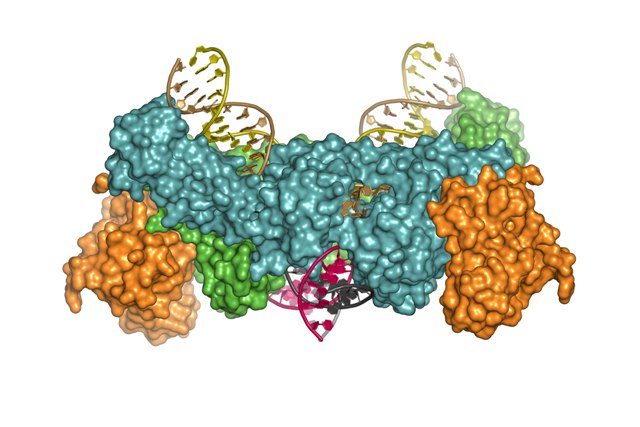
“It has truly been a breathtaking ride. Only 18 months ago we had a rather sketchy understanding of retroviral integration. Now we have obtained snapshots depicting the whole process in atomic detail. The new 3D structures capture the retroviral integration machine in action.”

Dr Peter Cherepanov, Imperial College London

These findings may enable improvements in gene therapy to correct gene malfunctions. A disabled version of a retrovirus can be used to insert a functional copy of a gene into a human chromosome. Although such approaches have proven quite successful, they are still experimental, requiring more work to make them safer.
 
Dr Cherepanov explains how his group’s latest findings could help: “One of the main problems with the current method is that retroviral integration is too random. While being very efficient, by its nature, integration is not very selective with respect to its target, the chromosomal DNA. Ideally, we want to insert therapeutic genes in pre-defined, safe locations of the human genome.
 
Using the new structural information we have uncovered, researchers can now start trying to tweak the retroviral integration machinery to make it more suitable for practical applications. For example, we might be able to design integrase that would be highly selective for target DNA sequence.”

The structural data collection was carried out on the I02 macromolecular crystallography experimental station at the Diamond synchrotron in Oxfordshire.

“This kind of fundamental research is vital if we are to advance our understanding of the viruses and diseases that affect millions of people around the world. Knowing the 3D structure of the mechanisms involved is like being able to see inside the engine of your car. If you can actually see what is happening, you get an idea of how you can fix it. At Diamond we produce the extremely intense X-ray beams required for looking at the molecular interactions involved in a variety of biological processes.”

Professor Thomas Sorensen, Diamond Light Source

Advances in structural biology have accelerated greatly as a result of access to the synchrotron facilities that have been developed around the world in the past 25 years. Biologists have been swift to recognise the huge potential that lies behind understanding the multitude of processes that take place within living organisms at a molecular level. Researchers in the UK are at the forefront of this work and Diamond Light Source plays its part in providing cutting edge facilities for protein structure determination. Diamond currently has five experimental stations dedicated to structural biology as well as an on-site membrane protein laboratory. Previous breakthroughs using structural data from Diamond include gaining a better understanding of hypertension in the pre-natal condition pre-eclampsia, learning how a key tuberculosis drug is activated, understanding how bird flu can affect humans, and solving the 3D atomic models of a single transporter protein responsible for the movement of essential chemicals into cells of the body.

The mechanism of retroviral integration through X-ray structures of its key intermediates, Goedele N. Maertens, Stephen Hare & Peter Cherepanov, Nature November 2010.
DOI 10.1038/nature09517

  • Contact Us
  • About Diamond Light Source
  • Careers
  • Procurement
  • Legal notices & Cookie policy
  • Supply Chain Transparency

Diamond Light Source

Diamond Light Source is the UK's national synchrotron science facility, located at the Harwell Science and Innovation Campus in Oxfordshire.

Copyright © 2020 Diamond Light Source


Diamond Light Source Ltd
Diamond House
Harwell Science & Innovation Campus
Didcot
Oxfordshire
OX11 0DE

See on Google Maps

Diamond Light Source® and the Diamond logo are registered trademarks of Diamond Light Source Ltd