High-energy X-rays produced by Diamond Light Source, the UK's national synchrotron science facility, have revealed the earliest rugose coral recorded to date. Using high-resolution tomography enabled by the Diamond synchrotron, researchers from Amgueddfa Cymru – National Museum Wales and Golestan University (Iran) were able to create 3D images of the rare fossil, without having to sample it destructively. The clear, detailed images confirmed that the fossil was a 462 million year-old rugose coral, five million years older than previous discoveries of this type. The results, published online in Geological Magazine*, demonstrate that this non-destructive synchrotron technique holds great promise for the "virtual dissecting" of fossils and could be set to transform palaeontological studies in the UK.
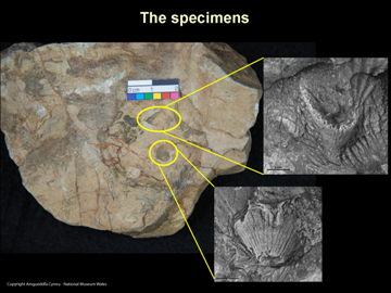
 Specimens of the fossilised coral. Image courtesy of Dr Christian Baars, Amgueddfa Cymru.
The bright, intense and finely focused X-rays produced by a synchrotron are expert at revealing the atomic and molecular details of tiny nano- or micrometre samples of material. But fossils tend to be giant in the world of synchrotrons, needing extremely high-energy X-rays to penetrate the millimetres of rock they are encased within. These recent results prove that the technique works. Synchrotron tomography is effectively simulating the traditional method of slicing sections from the fossil to help identify what it is and when it became fossilised. The advantage is that the fossil remains intact and the information uncovered is incredibly detailed. Current developments at the Diamond synchrotron are set to enable researchers to discover the chemical details of their fossil, along with their image.
Lead researcher, Dr Christian Baars, from Amgueddfa Cymru, will present the rugose coral 3D images on Monday 17th December 2012 at the Palaeontological Association’s Annual Meeting, which takes place at University College Dublin. Dr Baars explains, "Traditionally, in order to accurately describe corals and some other kinds of fossils, we would section and photograph or draw these fossils. Being a destructive technique, this is not always desirable but, until recently, has been unavoidable. X-ray tomography carried out at Diamond enabled us to produce images of these fossils non-destructively by measuring the relatively small density contrast between calcite and quartz to give high quality reconstructions of the fossil. It is extremely exciting to see that this technique works. It could change the way we carry out taxonomy of fossils. National Museum staff are already planning to analyse further samples, for instance the tiny fossilised eggs of 450 million year old sea creatures."

Morphology of rugose corals. Image courtesy of Dr Christian Baars, Amgueddfa Cymru.
Rugose corals are thought to have evolved during the Ordovician geological period (488 – 444 million years ago) from animals such as sea anemones that started secreting a hard skeleton made of calcite. Soft-bodied animals like sea anemones have a very poor fossil record as, normally, only the hard skeleton of animals is preserved. The new fossil is also of particular interest to palaeontologists because it lived in a part of the world which was in similar latitudes as today's central Argentina or southern Australia when most other early rugose corals lived at around the equator. This is important as it changes our ideas of how and where these organisms evolved.
Dr Robert Atwood, Senior Support Scientist on Diamond’s JEEP (Joint Engineering and Environmental and Processing) beamline adds, “This work has been really important for Diamond. It has enabled us to establish that we can examine partially silicified fossils, which are embedded in a calcareous rock matrix, and help palaeontologists uncover secrets that have been locked away for millions of years. The tomography that we can offer researchers provides good contrast because we provide a high energy, monochromatic X-ray beam which is very stable. This means the beam can penetrate relatively thick rock sections to observe the fossils that lie inside with good sensitivity to small differences in the minerals. This helps researchers like Dr Baars avoid cutting rare and unique specimens into pieces. In the case of the rugose coral, we rotated the fossil in the X-ray beam and obtained 6,000 images during the experiment.”
Dr Atwood continues, “Using our post-experimental data analysis, we were then able to then put these images together and produce the three-dimensional reconstruction of the fossil. This reconstruction gave Dr Christian Baars and his colleagues the clues they needed to confirm the fossil as the earliest rugose coral recorded to date. We are looking forward to working with other museums and university based palaeontologists to help them gain access to this exciting synchrotron technique.”
*The earliest rugose coral.'
Christian Baars, Mansoureh Ghobadi Pour & Robert Atwood
 Read the Museum of Wales article "Ancient fossil meets modern technology"
![]()
![]()
 Specimens of the fossilised coral. Image courtesy of Dr Christian Baars, Amgueddfa Cymru.
 Read the Museum of Wales article "Ancient fossil meets modern technology"