The Magnetic Materials Group comprises scientists and engineers working across five beamlines (I06, I10, I16, B16 and I21) covering the soft to hard X-ray range, and a variety of techniques, from resonant X-ray scattering, coherent diffraction imaging, photoemission electron microscopy to absorption spectroscopy. Over the last year, our research community has explored the fundamental properties of a wide range of naturally occurring and fabricated materials to discover a wealth of fascinating new phenomena. In this contribution, we present research from the beamlines demonstrating how X-ray diffraction and imaging can isolate surface effects in chiral spin structures, and how confined volumes can dramatically enhance the growth of single-crystals. We also present the development of a new form of non-linear X-ray spectroscopy, which has enormous potential as an atomic probe of valence states. The results, as a whole, highlight the need for improved theoretical models to underpin technique and material development.
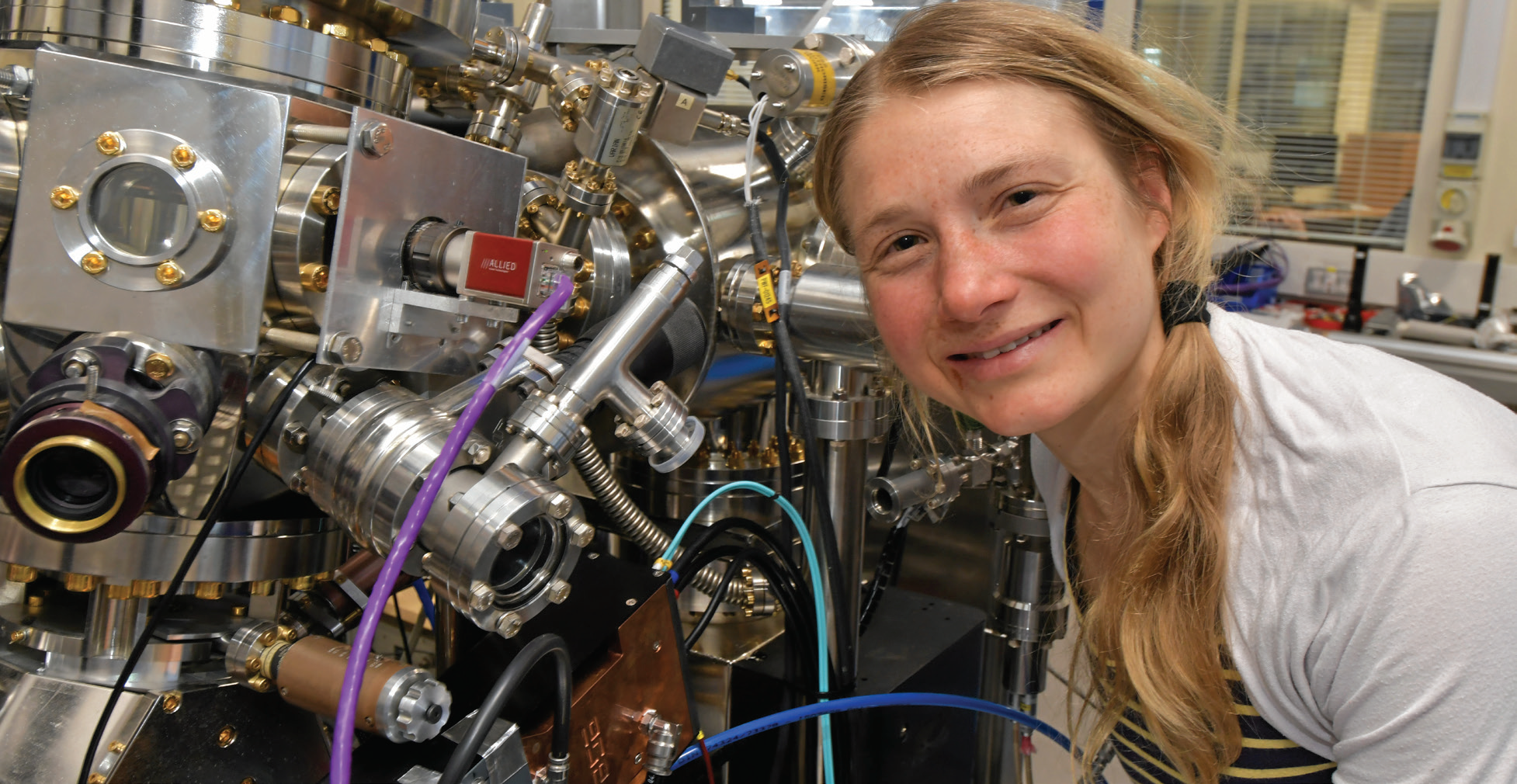
- Sonka Reimers, a joint Nottingham and Diamond PhD student, using I06 to investigate how to store and read information in antiferromagnets.
Vortices are naturally occurring structures that form on cosmic as well as microscopic length scales. On the other hand, vortices in magnetic materials are rare since they are generally destroyed by long-range interactions. On beamline I06, researchers have discovered such structures in antiferromagnetic (AF) thin films of iron oxide (Fe2O3) for which the destructive, long-range interactions are absent due to the lack of a net magnetic moment. Using X-ray Magnetic Linear Dichroism (XMLD) based high-resolution vector magnet imaging in the Photoemission Electron Microscope (PEEM), vortex-antivortex pairs have been discovered and their interactions with a ferromagnetic cobalt overlayer determined. Furthermore, since Fe2O3 exhibits a spin flop transition, which aligns the magnetic moments along the c-axis, it naturally provides new opportunities to test theoretical models of chiral structures formed by nucleation rather than fluctuations. The I06 PEEM has also been used by researchers to understand how AF domain wall motion can depend on the polarity of an injected current (see Fig. 1). In previous work, orthogonal in-plane current pulses were used to induce 90o rotations of AF domains that were not polarity dependent. In the new work, researchers have demonstrated that AF domain walls can be manipulated to realise stable and reproducible domain changes using just two electrical contacts. This represents a major breakthrough in switching AF materials since the effect can occur at much lower current densities than those needed for coherent domain rotation.
Over the last year, our research community has explored the fundamental properties of a wide range of naturally occurring and fabricated materials to discover a wealth of fascinating new phenomena.
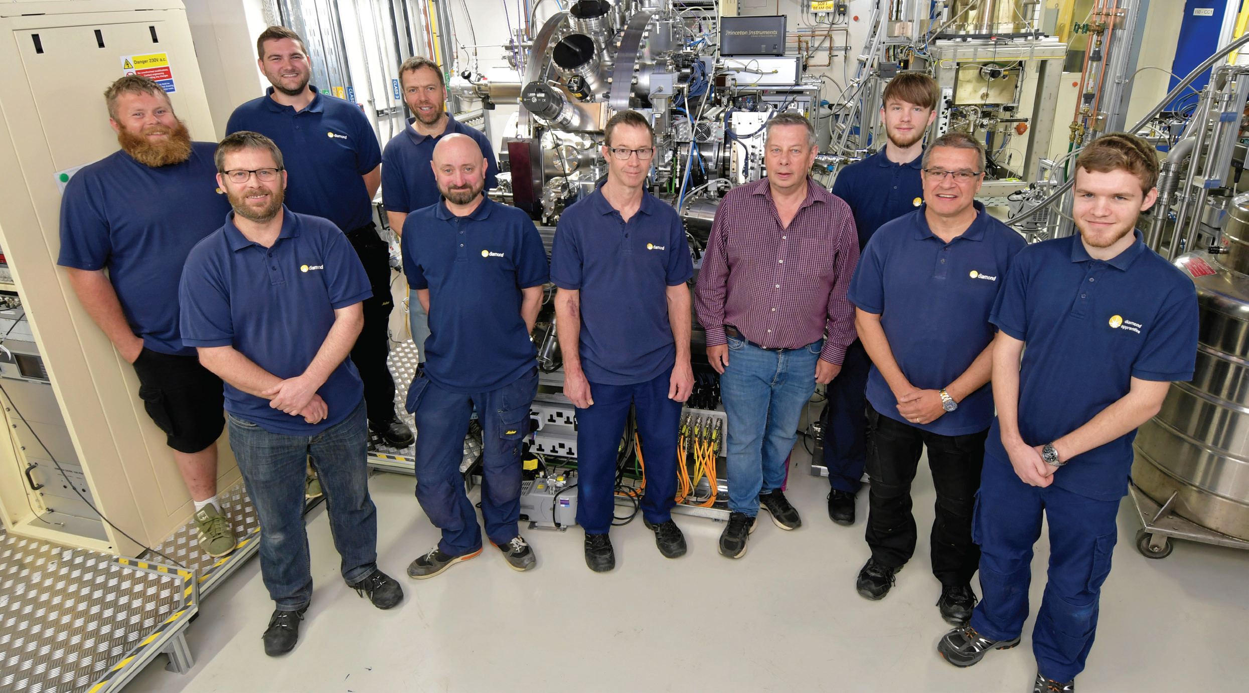
- The Magnetic Materials Group Mechanical & Electrical Technicians that underpin the research across the facilities of the group, from left to right, back: Tom Rice, Chris Callaway, Andy Malandain, Ryan Russell. Front: Matthew Hilliard, Lee White, Mark Sussmuth, Richard Mott, Mike Matthews, Sam Embling.
Skyrmions are topologically protected spin textures that arise from a subtle balance between the energies associated with the exchange interaction, the spin-orbit interaction and magnetic anisotropy. Generally speaking, there are two types of skyrmions (Néel and Bloch), but recent theoretical work has predicted a chiral twist to this story at the surface of a material. By utilising the unique properties of the RASOR diffractometer on beamline I10 researchers have revealed how skyrmion structures change close to the surface using extinction effects in polarised soft X-ray resonant elastic scattering. The magnitude of these distortions is much larger than predicted by theory, indicating the need to include surface effects more accurately in theoretical models.
Spectroscopic probes of semiconductor band gaps or phenomena associated with states close to the Fermi energy have had a rich history of development at optical wavelengths. On beamline I16 researchers have combined the highly monochromatic and collimated X-ray beam with a carefully designed experimental setup to detect Parametric Down Conversion (PDC) of X-ray photons at energies corresponding to optical photons. The importance of PDC is that it detaches the spatial resolution limit from the wavelength of the probe light used. The breakthrough on I16 means that, for instance, local valence-electron charge maps can be generated using the sensitivity of visible light spectroscopy, but with atomic scale resolution. Moreover, numerical simulations predict that the PDC effect is two orders of magnitude weaker than that measured in the experiment, implying significant opportunities for a more detailed theoretical understanding of resonant and non-resonant effects in non-linear X-ray spectroscopy.
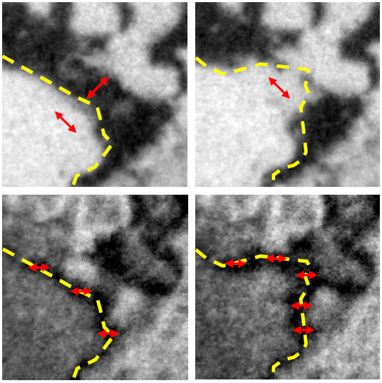
- Figure 1: XMLD-PEEM images showing AF domain rotation and domain wall motion after current injection into a CuMnAs film grown on GaP. (Top) sample oriented to show AF domains before (left) and after (right) current injection. (Bottom) sample oriented to show the corresponding AF domain wall before (left) and after (right) current injection. The field of view is 4 μm. The red arrows indicate the local AF axis and the broken yellow lines indicate the position of the AF domain wall. Adapted from P. Wadley et al., Nat. Nanotech. 13, 362 (2018).
The Magnetic Materials Group has had a busy year improving and extending the capabilities of its facilities for X-ray research. I21 accepted first users in October 2017 and has been operating in optimisation mode with ~50% of available beamtime allocated to the user programme. Towards the end of 2018, the beamline spectrometer was transformed from a discrete-port setup to a continuous-rotation configuration allowing, amongst other things, studies of emergent phenomena throughout the Brillouin zone. The exceptional efficiency of I21 has meant that the user community, together with the beamline team, have been able to measure magnon dispersion at the O K-edge, detect extremely weak signals from thin films of only a few unit cells, and resolve f-f excitations in heavy fermion systems. On I16, a new polarisation analyser stage and detector manifold has been installed, along with a Pilatus in vacuum area detector allowing improved efficiency at photon energies down to the S K-edge (~2.5 keV). On I10, a new feedback system allows improved spatial resolution when mapping, for instance, electronic and magnetic domain structures. The new 1.6T electromagnet end station is now in the commissioning phase and will enable much faster absorption spectroscopy studies complementing the existing 14T superconducting facility. A new direct-electron detector has been installed on the I06 PEEM improving the quantum efficiency and increasing the frame rate to >1000 fps. The PEEM manipulator has also been upgraded to reach lower temperatures (<60 K) whilst maintaining rapid azimuthal rotation of the sample. On B16, an X-ray imaging system has been developed and combined with on-the-fly scanning tomography enabling data acquisition times to be reduced from hours to seconds using the intense white beam available. In addition, a new Merlin quad-detector chip, based on the Medipix3, has been added, which further increases the efficiency of the diffraction and imaging facilities of the beamline. The remarkable imaging capabilities of B16 are demonstrated by recent work on the formation of aragonite in confined membranes. Using commercial filtration membranes, researchers were able to determine that precipitation of single-crystal aragonite in 25 nm diameter membranes could be significantly enhanced in the presence of Sulphur and Magnesium ions.
The Magnetic Materials Group is dedicated to continually developing and improving its facilities and off-line laboratories and would welcome further input from our user community. We organise regular workshops to explore new scientific and technical opportunities together with our user community. In the past year we have run workshops on Resonant Inelastic and Elastic X-ray Scattering (June 2018), Opportunities for Quantum Materials Research at Diamond-II (September 2018) and a workshop for the new Aberration- Corrected PEEM at Diamond (January 2019). Our objective is to run a suite of state-of-the-art polarised X-ray beamlines with leading edge software, along with access to advanced on-site sample characterisation facilities.

Diamond Light Source is the UK's national synchrotron science facility, located at the Harwell Science and Innovation Campus in Oxfordshire.
Diamond Light Source Ltd
Diamond House
Harwell Science & Innovation Campus
Didcot
Oxfordshire
OX11 0DE
Copyright © Diamond Light Source. Diamond Light Source® and the Diamond logo are registered trademarks of Diamond Light Source Ltd
Registered in England and Wales at Diamond House, Harwell Science and Innovation Campus, Didcot, Oxfordshire, OX11 0DE, United Kingdom. Company number: 4375679. VAT number: 287 461 957. Economic Operators Registration and Identification (EORI) number: GB287461957003.