Diamond Home Page
  • Diamond Home Page
  • About Us
  • For Users
  • Industry
  • Public
  • Science
  • Instruments
  • Careers
  • More
Search

On this website

About Diamond

  • About Diamond
  • About Synchrotron
  • News and Features
  • Events
  • Contact Us
  • FAQ

For Users

  • Apply for beamtime
  • User guide
  • Diamond Users Commitee
  • FAQ

Industry

  • Techniques Available
  • Industry Research
  • Industry Case Studies
  • News
  • Meet the Industry Team

Science

  • Research
  • Computing
  • The Machine
  • Publications
  • Research Expertise
  • Membrane Protein Laboratory
  • Additional Facilities

Instruments

  • All the Instruments
  • Macromolecular Crystallography
  • Soft Condensed Matter
  • Imaging and Microscopy
  • Biological Cryo-Imaging
  • Magnetic Materials
  • Structures and Surfaces
  • Crystallography
  • Spectroscopy

Careers

  • Vacancies
  • Info for applicants
  • Company Benefits
  • Apprenticeships
  • PhD Studentships
  • Work Placement

Public

  • News
  • How Diamond Works
  • Multimedia
  • Features
  • Visits Us
  • For School

Procurement

  • Non-OJEU Tender Notices
  • OJEU PINs
  • OJEU Tender Notices
  • Registration Form

Software & Tools

  • Publications Database
  • iSpyB
  • User Administration System

Main Content

A brighter light for science
Sub-navigation
  • About Us
  • Governance
  • Legal & Compliance
  • News & Literature
  • Events
  • Contact Us
  • Location

In This Section

Sub Navigation
  • News & Science Highlights
  • Annual Review
    • Diamond Annual Review 2019
      • Foreword
      • CEO Welcome
      • Beamline Development and Technical Summary
      • Science Group Updates
        • Macromolecular Crystallography Group
        • Biological Cryo-Imaging Group
        • Structures and Surfaces Group
        • Magnetic Materials Group
          • Sarnjeet Dhesi, Science Group Leader
          • Magnetic knots suggest a pathway to energy-efficient computers
          • Next-gen memory: How skyrmions behave at surfaces
          • Nonlinear spectroscopy with X-rays
          • Confinement effects promote the formation of aragonite
        • Imaging and Microscopy Group
        • Crystallography Group
        • Spectroscopy Group
        • Soft Condensed Matter Group
      • Phase III Overview
      • Integrated Facilities
      • Collaborations
      • Machine Operation and Development
      • Optics and Metrology Group
      • Detector Group
      • Scientific Software, Controls and Computation
      • Key Facts and Figures
      • Industrial Liaison at Diamond
      • Engaging with Diamond Light Source
      • Governance and Management
      • Committee Membership
    • Diamond Annual Review 2018
    • Diamond Annual Review 2017
    • Diamond Annual Review 2016
    • Diamond Annual Review 2015
    • Submission Guidelines
  • Literature Reviews
  • Impact Case Studies
  • Print Publications
  • Press & Media

Opportunities at Diamond

Learn more about career and student opportunities at Diamond:

Vacancies

Students

Subscribe to our Corporate and Science Update mailing list

Keep up to date with the latest research and developments from Diamond. Sign up for news on our scientific output, facility updates and plans for the future.

Subscribe here

  1. Diamond Light Source
  2. News & Literature
  3. Annual Review
  4. Diamond Annual Review 2019
  5. Science Group Updates
  6. Magnetic Materials Group
  7. Nonlinear spectroscopy with X-rays

Nonlinear spectroscopy with X-rays

Related publication: Schori A., Bömer C., Borodin D., Collins S. P., Detlefs B., Moretti Sala M., Yudovich S. & Shwartz S. Parametric Down- Conversion of X Rays into the Optical Regime. Phys. Rev. Lett. 119, 253902 (2017). DOI: 10.1103/PhysRevLett.119.253902
 
Publication keywords: Spectroscopy; Nonlinear optics; Second order nonlinear optical processes; Parametric down-conversion; Valence electrons

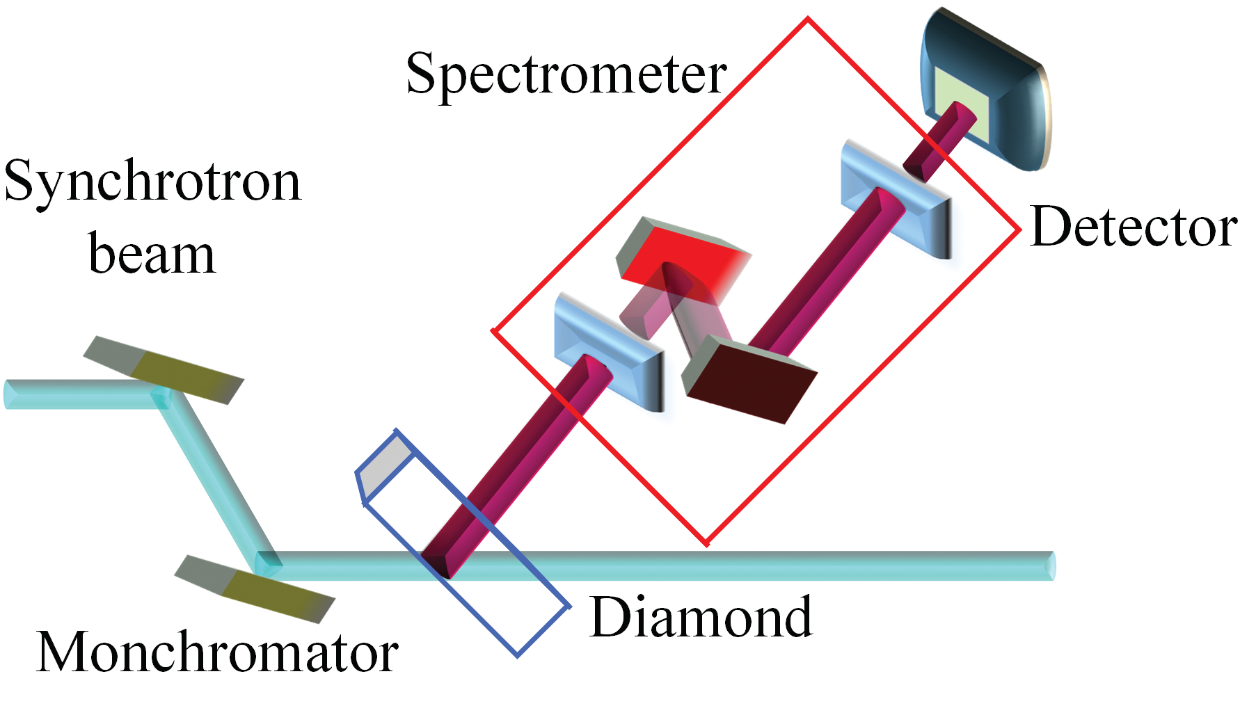
An international team of researchers has demonstrated a novel method for studying the microscopic structure of chemical bonds, the valence electron density of crystals, and light-matter interactions at the atomic scale resolution, with synchrotron radiation. The goal of the work was to develop a technique that provides spectroscopic and structural information on valence electrons in a single measurement, this new approach enables the measurements of atomic scale interactions of valence electrons, providing access to microscopic scale effects.

The team chose the Materials and Magnetism beamline (I16) at Diamond Light Source for their experiments due to its high photon flux, the high collimation of the X-ray source, and the combined energy resolution of the monochromator-analyser setup. They combined spectroscopic information with structural information to enhance understanding of microscopic inter-molecular processes.
Their results provide the first observation of parametrically down-converted (PDC) X-ray signal photons at photon energies that correspond to idler photons at optical wavelengths. They demonstrate that the conversion of X-rays into optical photons may be used as a new tool to probe microscopic valence charge densities and optical properties of materials on the atomic scale with synchrotron radiation. This novel tool can be used to test and improve the understanding of condensed matter physics. 
Figure 1: Experimental setup. The scattering plane of the analyser was normal to the scattering plane of the sample. S1 and S2 are the slits before the analyser and before the detector. The analyser
<br/>was a Si(440) crystal and the detector was an avalanche photodiode.
Figure 1: Experimental setup. The scattering plane of the analyser was normal to the scattering plane of the sample. S1 and S2 are the slits before the analyser and before the detector. The analyser
was a Si(440) crystal and the detector was an avalanche photodiode.

The understanding of the interactions of valence electrons with light, and with each other, is essential for the understanding of many phenomena in physics, chemistry, and biology. However, although optical radiation has been extensively used to study the properties of valence electrons, optical radiation spectroscopy methods do not provide atomic scale information due to their long wavelengths. While X-ray Bragg diffraction can reveal structural information at the atomic scale, those types of experiments cannot provide spectroscopic information on valence electrons. Thus, the understating of the microscopic nature of valence electron interactions remains a great challenge.

Nonlinear interactions of X-rays and long wavelengths can provide insight into the microscopic structure of chemical bonds, the valence electron density of crystals, and light-matter interactions at the atomic scale resolution1. In essence, the high resolution stems from the short wavelengths of X-rays, whereas the long wavelength field is used to probe the interactions with the valence electrons. The effect could be considered as X-ray diffraction from optically modulated charge densities. In analogy to Bragg diffraction, the atomic scale resolution can be achieved by measuring the intensities for different atomic planes but, in contrast to Bragg diffraction, in the nonlinear process the intensity is proportional to the Fourier coefficient of the valence electrons, and not to the coefficient of the core electrons.

To date, there was only one observation of nonlinear interactions between X-rays and optical radiation. Glover et al. measured nonlinear wave mixing between X-rays and optical radiation by using the first hard X-ray free-electron laser2, and an optical laser. However, it is very challenging to perform further progress with this effect, mainly because it requires high optical intensities, which are above the radiation damage threshold of most materials. 
Figure 2: (a) X-ray signal count rate as a function of the analyser detuning from the photon energy of the input beam. (b) X-ray count rate as a function of the pump deviation angle from the phase
<br/>matching angle. The blue dots are the experimental results and the solid red line is calculated from theory.
Figure 2: (a) X-ray signal count rate as a function of the analyser detuning from the photon energy of the input beam. (b) X-ray count rate as a function of the pump deviation angle from the phase
matching angle. The blue dots are the experimental results and the solid red line is calculated from theory.
In this work, we have demonstrated the nonlinear effect of parametric down-conversion (PDC) of X-rays into the optical regime. In this nonlinear effect, which is another wave mixing effect that relies on the same nonlinear mechanism, the input photons interact with vacuum fluctuations to generate correlated X-ray and optical photon pairs. Since the photons are always generated in pairs, and since the process conserves energy and momentum, the generation rate, the photon energy, and the angle of propagation of the generated X-ray photons are perfectly correlated with the long wavelength photons. Consequently, it is sufficient to measure only the X-ray signal to probe the properties of the sample at energies corresponding to long wavelength photons. Several demonstrations of PDC into extreme ultraviolet were reported3,4, but the extension of this method into the optical regime is more challenging due to the proximity of the photon energy to the input photon energy.
 
The experiments were conducted at ID-20 (ESRF) and at I16. The measurements were performed with a highly collimated monochromatic beam at 9 and 11 keV, to illuminate a diamond crystal for the generation of the PDC. The emitted X-ray photons were collected by a system that contains slits, a crystal analyser, and a detector (Fig. 1). The diffraction was from the C(220) atomic planes.
 
Fig. 2a shows the spectrum of the effect. The peak on the left corresponds to the elastic scattering, and the broad peak is the PDC signal. The peak is observed at 7.1 eV, where the efficiency of the PDC is the largest. This energy is near the bandgap of the diamond crystal, where the density of states of the valence electrons is the highest. This observation demonstrates the ability to measure the valence electron spectral dependencies.
 
To demonstrate the ability to calculate the Fourier component of the nonlinear susceptibility, the rocking curve of X-ray signal of PDC at an optical wavelength that corresponds to 2.2 eV was measured (Fig. 2b). The small peak on the left is the residual elastic, and the peak centred at 15 mdeg from the origin is the PDC signal. The Fourier coefficient of the susceptibility obtained from the measurement is comparable to the theoretical prediction, and to the result of X-ray and optical mixing experiment2.
 
The first observation of PDC of X-ray into optical radiation is reported. The results advance the possibility to use the effect as a new tool to probe microscopic valence charge densities, and optical properties of materials on the atomic scale. This novel tool can be used to test and improve the understanding of condensed matter physics. 
 

 References:

  1. Freund I. et al. Optically modulated X-ray diffraction. Phys. Rev. Lett. 25, 1241–1245 (1970). DOI: 10.1103/PhysRevLett.25.1241
  2. Tamasaku K. et al. Visualizing the local optical response to extreme-ultraviolet radiation with a resolution of I λ/380. Nat. Phys. 7, 705–708 (2011). DOI: 10.1038/nphys2044
  3. Hastings J. B. et al. X-ray and optical wave mixing. Nature 488, 603–608 (2012). DOI: 10.1038/nature11340
  4. Borodin D. et al. High energy-resolution measurements of x-ray into ultraviolet parametric down-conversion with an x-ray tube source. Appl. Phys. Lett. 110, 131101 (2017). DOI: 10.1063/1.4979413
 Funding acknowledgement:
Israel Science Foundation (ISF) (1038/13); Marie Curie FP7 Integration Grant within the 7th European Union Framework Programme (PCIG13- GA-2013-618118); Ministry of Aliyah and Immigrant Absorption of Israel; Deutsche Forschungsgemeinschaft (DFG) via SFB925 (Teilprojekt A4).
Corresponding author:
Dr Sharon Shwartz, Bar-Ilan University, sharon.shwartz@biu.ac.il
  • Contact Us
  • About Diamond Light Source
  • Careers
  • Procurement
  • Legal notices & Cookie policy
  • Supply Chain Transparency

Diamond Light Source

Diamond Light Source is the UK's national synchrotron science facility, located at the Harwell Science and Innovation Campus in Oxfordshire.

Copyright © 2022 Diamond Light Source


Diamond Light Source Ltd
Diamond House
Harwell Science & Innovation Campus
Didcot
Oxfordshire
OX11 0DE

See on Google Maps

Diamond Light Source® and the Diamond logo are registered trademarks of Diamond Light Source Ltd

Registered in England and Wales at Diamond House, Harwell Science and Innovation Campus, Didcot, Oxfordshire, OX11 0DE, United Kingdom. Company number: 4375679. VAT number: 287 461 957. Economic Operators Registration and Identification (EORI) number: GB287461957003.

feedback