Diamond Home Page
  • Diamond Home Page
  • About Us
  • For Users
  • Industry
  • Public
  • Science
  • Instruments
  • Careers
  • More
Search

On this website

About Diamond

  • About Diamond
  • About Synchrotron
  • News and Features
  • Events
  • Contact Us
  • FAQ

For Users

  • Apply for beamtime
  • User guide
  • Diamond Users Commitee
  • FAQ

Industry

  • Techniques Available
  • Industry Research
  • Industry Case Studies
  • News
  • Meet the Industry Team

Science

  • Research
  • Computing
  • The Machine
  • Publications
  • Research Expertise
  • Membrane Protein Laboratory
  • Additional Facilities

Instruments

  • All the Instruments
  • Macromolecular Crystallography
  • Soft Condensed Matter
  • Imaging and Microscopy
  • Biological Cryo-Imaging
  • Magnetic Materials
  • Structures and Surfaces
  • Crystallography
  • Spectroscopy

Careers

  • Vacancies
  • Info for applicants
  • Company Benefits
  • Apprenticeships
  • PhD Studentships
  • Work Placement

Public

  • News
  • How Diamond Works
  • Multimedia
  • Features
  • Visits Us
  • For School

Procurement

  • Non-OJEU Tender Notices
  • OJEU PINs
  • OJEU Tender Notices
  • Registration Form

Software & Tools

  • Publications Database
  • iSpyB
  • User Administration System

Main Content

A brighter light for science
Sub-navigation
  • About Us
  • Governance
  • Legal & Compliance
  • News & Literature
  • Events
  • Contact Us
  • Location

In This Section

Sub Navigation
  • News & Science Highlights
  • Annual Review
    • Diamond Annual Review 2019
      • Foreword
      • CEO Welcome
      • Beamline Development and Technical Summary
      • Science Group Updates
        • Macromolecular Crystallography Group
          • Dave Hall, Science Group Leader
          • Using structure-based design to engineer chimeric protein vaccines
          • Understanding human sleeping sickness
          • Turning the plastic tide – engineering enzymes to tackle plastic pollution
          • Insc:LGN tetramers revert breast cancer stem cell expansion by promoting asymmetric cell divisions
          • Toward remote control of an ‘undruggable’ dynamic enzyme with structurebased fragment screening
          • Parkinson’s: Completing the Parkin activation puzzle
        • Biological Cryo-Imaging Group
        • Structures and Surfaces Group
        • Magnetic Materials Group
        • Imaging and Microscopy Group
        • Crystallography Group
        • Spectroscopy Group
        • Soft Condensed Matter Group
      • Phase III Overview
      • Integrated Facilities
      • Collaborations
      • Machine Operation and Development
      • Optics and Metrology Group
      • Detector Group
      • Scientific Software, Controls and Computation
      • Key Facts and Figures
      • Industrial Liaison at Diamond
      • Engaging with Diamond Light Source
      • Governance and Management
      • Committee Membership
    • Diamond Annual Review 2018
    • Diamond Annual Review 2017
    • Diamond Annual Review 2016
    • Diamond Annual Review 2015
    • Submission Guidelines
  • Literature Reviews
  • Impact Case Studies
  • Print Publications
  • Press & Media

Opportunities at Diamond

Learn more about career and student opportunities at Diamond:

Vacancies

Students

Subscribe to our Corporate and Science Update mailing list

Keep up to date with the latest research and developments from Diamond. Sign up for news on our scientific output, facility updates and plans for the future.

Subscribe here

  1. Diamond Light Source
  2. News & Literature
  3. Annual Review
  4. Diamond Annual Review 2019
  5. Science Group Updates
  6. Macromolecular Crystallography Group
  7. Understanding human sleeping sickness

Understanding human sleeping sickness

Related publication: Zoll S., Lane-Serff H., Mehmood S., Schneider J., Robinson C. V, Carrington M. & Higgins M. K. The structure of serum resistance-associated protein and its implications for human African trypanosomiasis. Nat. Microbiol. 3, 295–301 (2018). DOI: 10.1038/s41564-017-0085-3

Publication keywords: African trypanosomes; Innate immunity; Apolipoprotein L1; SRA

The worst forms of human sleeping sickness (African trypanosomiasis) are caused by a species of trypanosome (single-celled parasite) known as T. b. rhodesiense. Most African trypanosomes are unable to infect humans as they are killed by a component of human blood, known as trypanolytic factor, and only two trypanosome subspecies are able to infect humans. T. b. rhodesiense is able to do so due to the presence of a single molecule called SRA. Although it was clear that SRA is the sole molecule responsible for allowing this parasite to develop human resistance, we did not know how it worked, or what it looked like. We also did not know how it binds to the active component of the trypanolytic factor, a molecule called ApoLI.
 
A team of researchers from the Universities of Oxford and Cambridge investigated how SRA stops the trypanolytic factor from working, using the Macromolecular Crystallography beamlines I03 and I04-1 to collect diffraction data from crystals of SRA bound to an antibody, and to determine the structure of the molecule. They were able to work out what SRA looks like, and to map onto the structure the regions of SRA that bind to the trypanolytic factor. This provides a first view of how SRA gives a trypanosome the ability to infect humans. However, it still leaves many questions, and understanding exactly how ApoLI interacts with SRA remains an exciting future research challenge.
 

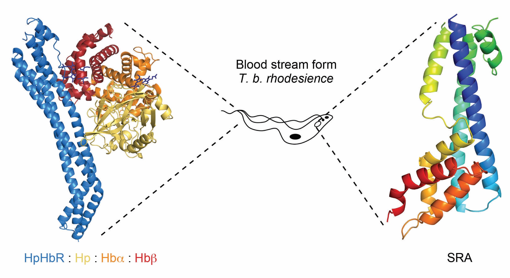
African trypanosomes are single-celled eukaryotic pathogens that are transmitted by tsetse flies, and live within the blood streams and tissue spaces of infected mammals. Infection causes disease, including the debilitating wasting illness of cattle, nagana, which restricts the productivity of livestock in sub-Saharan Africa. While there are numerous species of African trypanosome, only two subspecies, T. b. gambiense and T. b. rhodesiense, can proliferate in humans. This is due to lytic factors, present only in humans and some other primates, which are taken up by trypanosomes and causes their death1. Human-infective species are able to negate the action of this lytic factor. This confers the ability to grow within infected humans, and to cause the disease Human African trypanosomiasis, also known as sleeping sickness. The aim of our work is to understand how human-infective trypanosomes inactivate the lytic factor, and then to determine whether we can make them susceptible to lytic factor mediated killing.

Our first contribution towards these goals used beamline facilities at Diamond Light Source to understand how lytic factor is taken up by trypanosomes. The lytic factor is a complex of several components, including the pore-forming protein, apolipoprotein LI (ApoLI), which acts as the toxin and a complex of haptoglobin related-protein bound to haemoglobin (HprHb). Trypanosomes have a receptor on their cell surface that binds to haptoglobin-haemoglobin (HpHb), allowing them to scavenge this valuable nutrient from the blood. However, the presence of HprHb in lytic factor allows this toxin to hitch a ride on the receptor, taking it into the trypanosome cell, where it mediates its toxic effect. We used beamlines I03 and I04-1 to solve structures of HpHb receptors from human-infective and non-infective trypanosomes, alone and also bound to HpHb2-4. This revealed how the lytic factor is recognised by trypanosomes. It also helped us to understand how T. b. gambiense becomes human-infective, as it has a mutation of the HpHb receptor which reduces lytic factor uptake. Our structures revealed how this mutation decreases the affinity of the receptor for lytic factor, and how it helps to protect this trypanosome subspecies from destruction2,3.

We more recently turned our attention to T. b. rhodesiense, which counteracts the action of lytic factor by expressing an anti-toxin that binds to, and inactivates, ApoL1. The anti-toxin, known as the serum resistance associated protein (SRA), is found primarily within the lysosome of trypanosomes, and is sufficient to allow a trypanosome to become human infective. Our goal was to determine the structure of SRA, and to investigate how it binds to, and inactivates, ApoLI.
 
Determining the structure of SRA proved challenging. As we later discovered, a large loop at one end of SRA is flexible, and prevented the formation of well-ordered crystals. We were able to solve this problem by producing a panel of monoclonal antibodies that bind to SRA, and discovered that the complex of SRA with one of these antibodies formed crystals of sufficient quality5. Armed with data collected from beamline I03 at Diamond, we were then able to determine the structure of SRA. Together with data from hydrogen-deuterium exchange mass spectrometry, and validated by mutagenesis, this allowed us to map onto SRA the residues that interact with ApoLI.
 
This provided the first detailed structural insight into SRA and its mode of interaction with ApoLI5. It also revealed that prevailing models for how SRA prevents ApoLI function were not compatible with our structural findings. However, it still leaves many questions, and understanding exactly how ApoLI forms pores, and how the binding of SRA prevents these pores from forming, remains an exciting future research challenge.
Figure: The centre of the Figure shows a schematic of a blood stream form T. b. brucei. The left-hand side of the figure shows the T. b. rhodesiense haptoglobin-haemoglobin receptor (HpHbR, blue) bound
<br/>to a complex of the SP domain of haptoglobin (Hp, yellow), the a-subunit of haemoglobin (Hba, orange) and the b-subunit of haemoglobin (Hbb, red). This is found on the cell surface, and within the
<br/>flagella pocket of trypanosomes. The right-hand panel shows the surface resistance associated protein (SRA, rainbow) of T. b. rhodesiense, which is found primarily in the lysosome of trypanosomes.
Figure: The centre of the Figure shows a schematic of a blood stream form T. b. brucei. The left-hand side of the figure shows the T. b. rhodesiense haptoglobin-haemoglobin receptor (HpHbR, blue) bound
to a complex of the SP domain of haptoglobin (Hp, yellow), the a-subunit of haemoglobin (Hba, orange) and the b-subunit of haemoglobin (Hbb, red). This is found on the cell surface, and within the
flagella pocket of trypanosomes. The right-hand panel shows the surface resistance associated protein (SRA, rainbow) of T. b. rhodesiense, which is found primarily in the lysosome of trypanosomes.

References:

  1. Pays E. et al. The molecular arms race between African trypanosomes and humans. Nat. Rev. Microbiol. 12, 575 (2014). DOI: 10.1038/nrmicro3298.
  2. Higgins M. K. et al. Structure of the trypanosome haptoglobin{\ textendash}hemoglobin receptor and implications for nutrient uptake and innate immunity. Proc. Natl. Acad. Sci. 110, 1905–1910 (2013). DOI: 10.1073/pnas.1214943110
  3. Lane-Serff H. et al. Structural basis for ligand and innate immunity factor uptake by the trypanosome haptoglobin-haemoglobin receptor. Elife 3, e05553 (2014). DOI: 10.7554/eLife.05553
  4. Lane-Serff H. et al. Evolutionary diversification of the trypanosome haptoglobin-haemoglobin receptor from an ancestral haemoglobin receptor. Elife 5, e13044 (2016). DOI: 10.7554/eLife.13044
  5. Zoll S. et al. The structure of serum resistance-associated protein and its implications for human African trypanosomiasis. Nat. Microbiol. 3, 295–301 (2018). DOI: 10.1038/s41564-017-0085-3
Funding acknowledgement:
MRC (MR/L008246/1 and MR/P001424/1); Wellcome Trust.
 
Corresponding authors:
Prof Matt Higgins, University of Oxford, matthew.higgins@bioch.ox.ac.uk and Prof Mark Carrington, University of Cambridge, mc115@cam.ac.uk
  • Contact Us
  • About Diamond Light Source
  • Careers
  • Procurement
  • Legal notices & Cookie policy
  • Supply Chain Transparency

Diamond Light Source

Diamond Light Source is the UK's national synchrotron science facility, located at the Harwell Science and Innovation Campus in Oxfordshire.

Copyright © 2022 Diamond Light Source


Diamond Light Source Ltd
Diamond House
Harwell Science & Innovation Campus
Didcot
Oxfordshire
OX11 0DE

See on Google Maps

Diamond Light Source® and the Diamond logo are registered trademarks of Diamond Light Source Ltd

Registered in England and Wales at Diamond House, Harwell Science and Innovation Campus, Didcot, Oxfordshire, OX11 0DE, United Kingdom. Company number: 4375679. VAT number: 287 461 957. Economic Operators Registration and Identification (EORI) number: GB287461957003.

feedback