The Imaging and Microscopy Group brings together eight experimental facilities (I08, J08, DIAD, I12, I13-1, I13-2, I14 and ePSIC) which use electrons and X-rays to image samples under different experimental conditions across a diverse range of length scales and time scales. Different contrast mechanisms allow for imaging of sample properties such as elemental composition, density and structure. This ability to extract image sample properties in minute detail lends itself to a wide range of scientific areas, from chemistry and catalysis to environmental science, materials science, biology, medicine and cultural heritage.
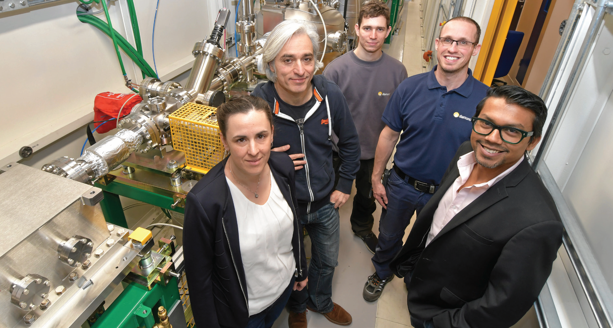
- DIAD beamline team, early 2019, left to right: Christina Reinhard, Michael Drakopoulos, Tom Yates, Pete Garland, Sharif Ahmed.
The Scanning X-ray Microscopy (SXM) beamline (I08) is for morphological, elemental and chemical speciation on a broad range of organic-inorganic interactions in a 250 - 4400 eV photon energy range, and sample investigations under ambient or cryogenic conditions. I08 has a range of applications including biological and biomedical sciences, earth and environmental science, geochemistry, and materials science. The main activity on I08 over the past year has been designing, constructing and testing various aspects of a new soft X-ray spectro- and tomo-ptychography branchline (J08). This new branchline is expected to be available for experiments in early 2020 and will provide spatial resolutions down to a few nm, providing a step change in imaging performance.
The Dual Imaging and Diffraction (DIAD) beamline will be the first beamline to offer two X-ray microscopy techniques (imaging and diffraction) applied synchronously with a switching time of 0.1s. This enables in situ structural characterisation experiments taking advantage of both techniques simultaneously. DIAD is being built to use light from a ten pole permanent magnet wiggler. The diffraction technique is conducted using monochromatic light, whereas the imaging technique can be performed with monochromatic or polychromatic (‘pink’) beam. The X-ray energy can be chosen separately for both techniques in the range from 8 - 38 keV. The beamline has completed construction of the Optics Hutch and took first light in December 2018. Next to a standard tomography setup, a mechanical test-rig for diffraction and tomography will be one of the main instruments to allow in situ experiments for a variety of scientific disciplines such as engineering and materials science, bio-materials and hard tissues, geology and mineralogy, and soil-plant interactions. The commissioning activities and construction of the experimental end station are ongoing with first users expected in early-mid 2020.
The Joint Engineering, Environmental and Processing (JEEP) beamline (I12) uses a 4.2 T superconducting wiggler to provide polychromatic and monochromatic X-rays in the energy range 50 - 150 keV. The high photon energies provide good penetration through large or dense samples. The beamline offers beam sizes ranging from 50 x 50 μm for diffraction, up to 90 x 25 mm for imaging. These beam characteristics enable the study of materials and processes inside sample environments without unacceptable attenuation of the beam, using macro-scale samples that are more representative of the process under study. X-ray techniques available are radiography, tomography, energy-dispersive diffraction, monochromatic and white-beam 2D diffraction/ scattering and small-angle X-ray scattering. The beamline’s two flexible experimental hutches allow users to bring their own rigs and sample chambers. I12 has a diverse user community (materials science and engineering; chemical processing; biomedical engineering; geoscience; environmental science; physics; palaeontology) who make full use of the beamline’s versatility. High-speed imaging and tomography has benefited from the integration of the Phantom Miro camera into the beamline data acquisition and processing pipelines. High-speed tomography of natural and artificial volcanic minerals during heating, cooling and compression at temperatures up to 1,300 °C have been undertaken by different groups, using this new capability, yielding insight into the development of gas bubbles and mineral precipitation during volcanic eruption. For diffraction and scattering measurements, a new Pilatus 2M Cd-Te detector has been commissioned and has entered user service, allowing faster and more sensitive detection of X-rays by photon-counting technology. The phase changes, strain and texture during friction welding of nickel alloys has been studied successfully with this method, enabling the changes at previously unreachable time-scales to be observed.
Different contrast mechanisms allow for imaging of sample properties such as elemental composition, density and structure.
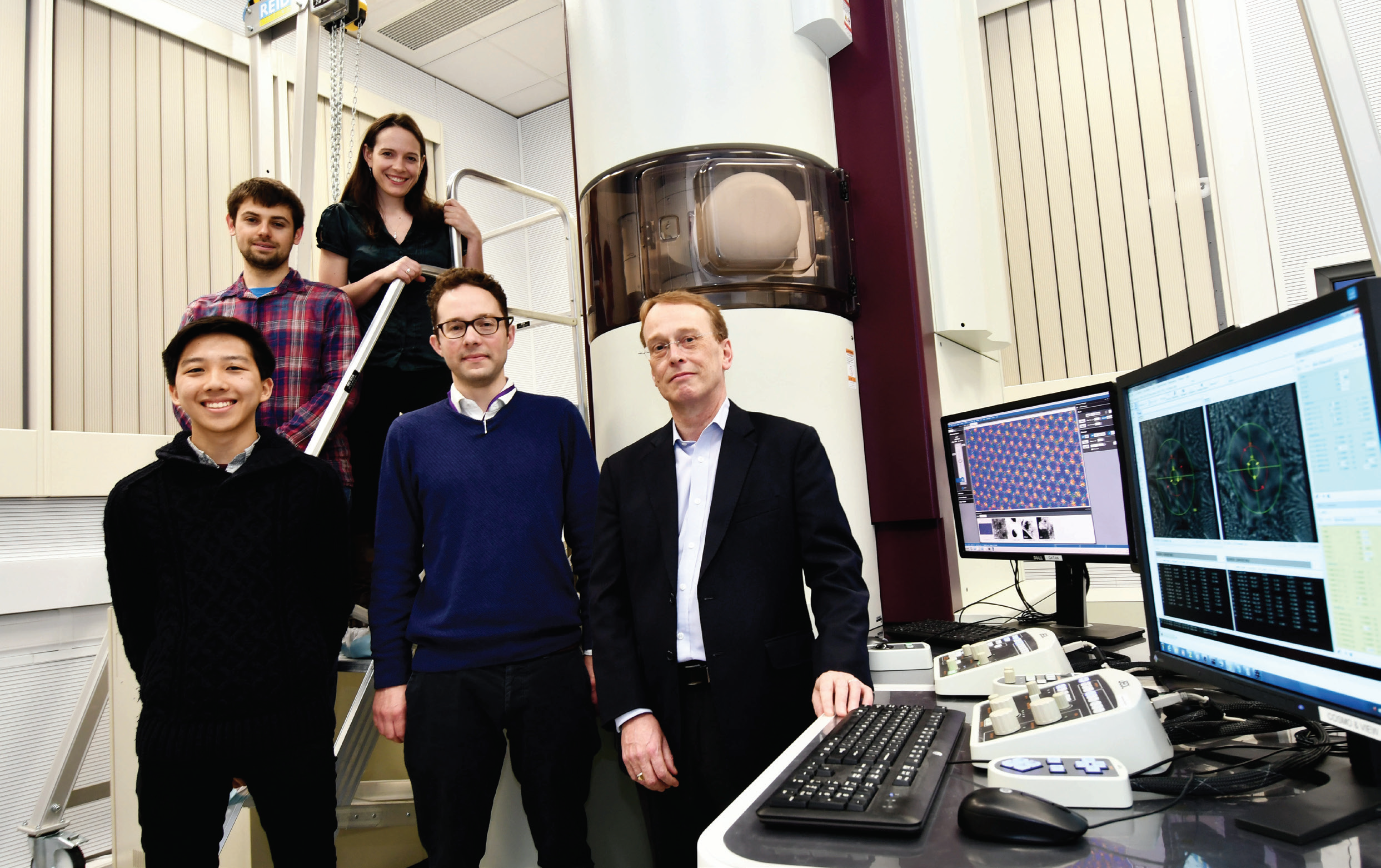
- ePSIC first users from the National Graphene Institute at The University of Manchester, left to right: Lan Nguyen, Aidan Rooney, Sarah Haigh, Manchester, with Christopher Allen and Angus Kirkland, Oxford.
The I13 Imaging and Coherence beamline is for multiscale imaging in the energy range of 6 - 30 keV. The achievable resolution ranges from several microns to some tens of nanometers with two branchlines operating independently for this purpose. The Diamond Manchester Imaging branchline performs mainly in-line phase contrast tomography with a strong emphasis on dedicated sample environments. A new full-field microscope using Zernike phase contrast imaging over a field of view of 50-100 μm and a resolution of 50 - 100 nm is now in operation, with a growing user community, allowing us to identify nano-sized structures under dynamic conditions. The highest spatial resolution, of 30 nm, is achieved on the coherence branch with ptychographic imaging. Continuous improvements such as new piezo stages and improved detector interfaces have reduced ptycho-tomography scans from days to a few hours, and ongoing fly-scanning developments aim to reduce this even further. Ptychography is now a routine standard user experiment and more advanced imaging modes such as 3D Bragg ptychography and simultaneous transmission and Bragg geometry measurements have been demonstrated and developed with users.
I14, the Hard X-ray Nanoprobe beamline, offers a small beam of 60 - 200 nm for high resolution imaging. I14 has entered its second year of operation and has developed and expanded its capabilities in X-ray fluorescence, diffraction and X-ray Absorption Near Edge Strucure (XANES) mapping. The integration of the Excalibur detector on I14 now enables diffraction data to be acquired at sampling times down to 10 ms. For XANES mapping, there have been a number of developments in automated drift correction to improve data quality. Corroded metal interfaces, metallic particles in cells and photovoltaic films are just a sample of the many science areas and successful experiments conducted. The beamline is still in its optimisation phase and new techniques and facilities such as ptychography are in development, and an increasing emphasis on in situ studies is driving a number of developments.
The electron Physical Science Imaging Centre (ePSIC) at Diamond consists of two transmission electron microscopes, a JEOL ARM 200 and a JEOL GRAND ARM 300, which were brought to Diamond through a collaboration with Johnson Matthey and the University of Oxford respectively. The ARM 200 is a state-of-the-art probe-corrected analytical microscope capable of atomic resolution electron energy loss and X-ray spectroscopy. The ARM 300 is a dedicated imaging instrument aligned across a wide range of accelerating voltages (30 - 300 keV). It is both probe- and imaging-corrected and has numerous detectors, including a fast direct electron detector (operating at up to 2000 fps). These combined capabilities make this a unique resource for electron microscopy within the UK. With in situ sample holders, users at ePSIC can perform variable temperature measurements from 100 to 1600 K to directly image the atomic structure of materials during thermally driven transitions. This in situ capability will be expanded upon over the coming year. An Oxford Instruments Energy Dispersive X-ray (EDX) detector has been added to the ARM 300 to allow combined X-ray spectroscopy and high-resolution imaging. The state of the art instrumentation available at ePSIC has attracted both established electron microscopists looking to develop new techniques, and scientists with limited previous electron microscopy experience interested in the atomic structure of their samples. The collaboration of the expert staff at ePSIC with this range of users is helping to bring cutting-edge microscopy techniques to the wider material science community. A science highlight from the last year has been the resolution of the structure of platelets in type 1a diamonds using the low voltage imaging capabilities of the ARM 300. The growing importance of ptychography has led to a project aimed at harmonising and improving the acquisition and analysis processes across the different X-ray and electron instruments.

Diamond Light Source is the UK's national synchrotron science facility, located at the Harwell Science and Innovation Campus in Oxfordshire.
Diamond Light Source Ltd
Diamond House
Harwell Science & Innovation Campus
Didcot
Oxfordshire
OX11 0DE
Copyright © Diamond Light Source. Diamond Light Source® and the Diamond logo are registered trademarks of Diamond Light Source Ltd
Registered in England and Wales at Diamond House, Harwell Science and Innovation Campus, Didcot, Oxfordshire, OX11 0DE, United Kingdom. Company number: 4375679. VAT number: 287 461 957. Economic Operators Registration and Identification (EORI) number: GB287461957003.