Dave Hall, Science Group Leader
Diamond provides a range of techniques for academic and industrial researchers studying the machines of life. As one of those techniques, Macromolecular Crystallography (MX) reveals the shape and arrangement of biological molecules at atomic resolution, knowledge of which provides a highly accurate insight into function. This can be combined with complementary information from many other techniques available at Diamond alongside lab based investigations to reveal the broader picture of molecular interactions and their effects.
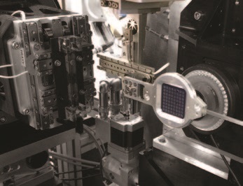
- Figure 1: The FT-SSX set-up available at I24.
MX is a core activity at Diamond with seven beamlines dedicated to the technique alongside the XFEL Hub, Membrane Protein Laboratory and XChemfragment screening facility for the extensive UK structural biology community as well as researchers in Europe and beyond. The staff of the MX group are recognised as innovative world leaders in MX, moving the goalposts of what is feasible for 'conventional' MX as well as developing techniques and beamlines that transform MX to the next level, enabling new experiments and methodologies. The group takes a long term approach to enabling new capabilities at its suite of beamlines to meet the current and future demands of an exacting community of scientists; in 2017 this was no different, as can be seen by the exciting developments here.
In 2017 the Long Wavelength MX beamline (I23) (dedicated to solving the crystallographic phase problem directly from native proteins) entered an exciting phase in its development, with a concerted push to facilitate user access alongside groundbreaking experiments with invited user groups. General user access to this technical tour de force was initiated in 2017 by its first call for users via the peer review proposal system for experiments in 2018 with great interest shown by the user community. Ongoing developments will continue on I23 to explore and exploit its potential to the full – we are entering a whole new world of what is possible with in vacuum MX and the samples and technology that enable this.
Serial Synchrotron Crystallography (SSX) is another emerging method driven, in part, by the sample requirements imposed by X-ray Free Electron Lasers (XFELs). Serial techniques can also be exploited at synchrotrons, especially at microfocus beamlines, opening up many new opportunities. A number of approaches including fixed targets and extruders have been tried and tested at Diamond’s Microfocus MX beamline (I24) and are available for general users. Expertise at I24 is centred on Fixed Target Serial Synchrotron Crystallography (FT-SSX). FT-SSX is attractive as it potentially offers high hit-rates coupled with modest sample consumption. Furthermore, the same approach can be used almost without modification at both synchrotrons and FELs.
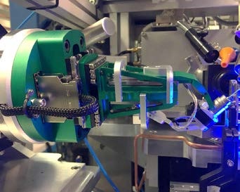
- Figure 2: The SmarGon goniometer installed in I04.
Our fixed target approach makes use of silicon nitride ‘chips’ with the current generation of chip capable of holding more than 25,000 crystals. Chips are mounted on a high-speed, high-precision xyz stage mounted at the sample position (Fig. 1). This setup allows data collection from all 25,600 positions on a chip in less than 10 minutes.
2017 has seen the final touches to the capabilities of the dedicated in situ VMXi beamline being commissioned and new workflows developed. The beamline is now at a state where regular user operation and further hardware/ software refinement is continuing in parallel. Input from initial users has been invaluable in progressing the beamline development, and indeed the demand for acting as beta testers from users has illustrated the keen interest amongst the MX community for the new capabilities that the beamline now provides. Alongside the beamline development, the VMXi team has also brought facilities together in a new central crystallisation laboratory located at the Research Complex (RCaH) next to Diamond. This facility is now open to users and can be used to prepare crystallisation experiments for the VMXi beamline – as well as for the Membrane Protein Laboratory and the XChem facility. Furthermore, the beamline team is working with the Diamond-based XFEL Hub to develop serial crystallography functionality which will also become part of the beamlines future offering to the MX community.
The last phase III MX beamline, VMXm, is progressing well. This beamline is designed to address the challenges of data collection from micron and sub-micron sized samples. With commissioning of the VMXm optics hutches essentially complete the end-station is now being assembled ready for commissioning and first experiments. First light into the experimental end-station hutch was seen in early March 2018 when work began on commissioning the microfocus optics. Offline assembly and testing of the sample environment is underway for incorporation into the end-station in April/May ready for first user experiments.
Our more conventional MX beamlines also continue to evolve to provide new functionality as well as increased performance. Through 2017 significant changes were made to the sample positioning systems on beamlines I03 and I04. Both are now equipped with a high precision, fast SmarGon multi-axis goniometer (Fig. 2) coupled to high capacity BART sample robots. The goniometers enable optimised data collection methods on these high throughput beamlines and have brought high speed snaked raster scanning, as developed for FT-SSX on I24, which enables the location of the smallest samples, best regions of samples and in automatic crystal X-ray centering methods. This latter use is part of a set of upgrades to all the MX cryogenic sample beamlines to bring automatic queued data collection to the user programme allowing expert systems to deliver the best data quality. The new goniometry set-up also allows I03 to switch to a fast, large format goniometer for in situ crystallisation plates for pathogenic samples or cornea experiments (Fig. 3). Standard MX in situ work is available on VMXi and I24.
Finally, fragment screening at the XChem facility attached to beamline I04- 1 is now fully integrated into Diamond’s user programme. Annually it hosts well over 30 screening experiments from academia and industry. In 2017, the programme contributed 35,000 of the more than 50,000 crystals shot at the beamline, but using less than one third of the total beamtime, illustrating the efficiency of the automated queue mode. With the programme consistently oversubscribed, and demand from industry growing, the facility is expanding its capacity: a dedicated user support team is being assembled, with extra capacity and an increase in dedicated lab space. Additional beamtime has been agreed, given or provided for in the allocation period for 2018
The progress made in 2017 for these facilities and their user programmes is underpinned by the dedication and expertise of all staff in the MX group working alongside support group staff from across Diamond as well as collaborations built across the world. The same staff engage in outreach, teaching and training for users, students and the public to build a greater understanding of the capabilities and science of MX in the broader setting of integrated structural biology.
- Figure 3: The large format goniometer in I03 supporting cornea lens experiments.

Diamond Light Source is the UK's national synchrotron science facility, located at the Harwell Science and Innovation Campus in Oxfordshire.
Diamond Light Source Ltd
Diamond House
Harwell Science & Innovation Campus
Didcot
Oxfordshire
OX11 0DE
Copyright © Diamond Light Source. Diamond Light Source® and the Diamond logo are registered trademarks of Diamond Light Source Ltd
Registered in England and Wales at Diamond House, Harwell Science and Innovation Campus, Didcot, Oxfordshire, OX11 0DE, United Kingdom. Company number: 4375679. VAT number: 287 461 957. Economic Operators Registration and Identification (EORI) number: GB287461957003.