 | ||
Making gold metamaterials with a twist |
Related publication:
Cseh L, Mang X, Zeng X, Liu F, Mehl GH, Ungar G, Siligardi G. Helically twisted chiral arrays of gold nanoparticles coated with a cholesterol mesogen. Journal of the American Chemical Society 137, 12736-12739, doi:10.1021/jacs.5b05059 (2015).
Keywords:
Liquid crystals; Nanoparticles; Circular dichroism; SAXS; GISAXS; Metamaterials; Plasmonic; Optical activity.
Nanometer sized metal particles can have unusual optical properties, with benefits that include potentially ‘cloaking’ objects rendering them invisible. Ordered arrays of gold or silver nanoparticles that can interact with circularly polarised (CP) light – light where the electric and magnetic fields rotate rather than vibrate – would extend potential application even further. CP light is used in many everyday objects, such as compact disc players and many mobile phone display screens. Future applications for CP light include optical computers, where light carries additional information via clockwise or anticlockwise spins. Methods of manipulating CP light are needed to enable such applications, and metamaterials (artificially constructed composite materials) with a twisted structure could potentially provide such manipulation. To make a twisted array of nanoparticles, cholesterol molecules were grafted onto the surface of gold nanoparticles. While X-ray diffraction experiments demonstrated that the resulting material was arranged in ordered arrays, it was unclear if the structure also had the crucial twist needed to manipulate CP light. Such a twist would show up in circular dichroism (CD) measurements, which track the difference between the absorption of right- and left-handed CP light in a material. Such a difference would exist if the structure was twisted. The Circular Dichroism beamline (B23) at Diamond Light Source provides a unique facility for measuring CD in small samples. Data from this beamline demonstrate that the combination of the gold nanoparticles with cholesterol did indeed have a helical twist. This new metamaterial opens up further optical applications for CP light.
Soft Condensed Matter Village & Materials Village | Beamlines B23/I22/B16
Near the frequency of plasmonic resonance in metal nanoparticles the refractive index becomes a complex quantity and its value can vary significantly close to the resonant frequency. If both the dielectric permittivity and magnetic permeability become negative, then the refractive index can also become negative, with the intriguing consequences mentioned above. It has been predicted theoretically that in order to alter the permitivity significantly and possibly even make it negative, relatively dense arrays of nanoparticles with periodicities much shorter than the wavelength of light are required1. Metal nanoparticles are normally coated with an organic layer to stop them from fusing together. By choosing rod- or disk-like liquid crystal forming molecules in the organic layer, anisotropic particle arrays can be obtained with different controllable particle packing modes2,3. Thus, the plasmonic band can be made to shift significantly as the material is rotated in a beam of linearly polarised light4.
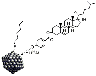
Figure 1: Gold nanoparticle with the two types of ligands attached.
The ultimate aim of this work is to investigate the possibility of developing plasmonic metamaterials that would interact with circularly polarised light, and that would distinguish between right- and left-handed CP light. The first step was to test if it was possible to obtain a dense ordered array of nanoparticles that would be not just anisotropic, but also spontaneously twisted. Gold nanoparticles, 2 nm in size on average, were therefore synthesised; they were initially coated with n-hexylthiol (Fig. 1). Subsequently about half the hexylthiols were replaced by a mesogenic, i.e. liquid crystal forming, ligand that contained cholesterol. As is well known, cholesterol is a chiral molecule, and a cholesterol ester was the first liquid crystal ever recognised as such; it forms the so called cholesteric, or chiral nematic LC phase, in which the molecular axis, or ‘director’, twists forming a helix.
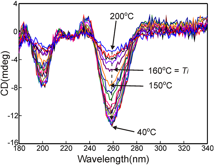
Figure 2: CD spectra in the UV region recorded at temperature intervals of 10 °C; Ti = 160 °C, coinciding with the largest change in CD.
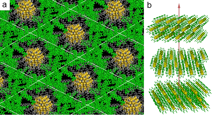
Figure 3: Structure of the twisted columnar liquid crystal phase of cholesterol-grafted gold nanoparticles: a) View along the gold strings (columns); the model was annealed by molecular dynamics; for clarity CPK rendering (yellow) is used for gold atoms, ball-andstick (green) for the cholesterol ester mesogens, and stick for the undecylthiol spacers and hexylthiol ligands; b) Schematic representation of the chiral columnar phase; colour code as in (a); the breaks in the stacks of columnar layers indicate that the helical pitch is long on the molecular scale.
X-ray diffraction is not very helpful when it comes to orientational rather than density fluctuations, and especially if these are on a >100 nm scale. In order to establish whether the superstructure of the LC phase is chiral, it was therefore necessary to use an optical technique. Thus circular dichroism was chosen, and it had to be measured as a function of temperature. Because the cholesterol ligand is intrinsically chiral, it was expected that the isotropic liquid would have a weak but finite CD signal. However, if the LC superstructure was itself chiral, then it would be expected to give a considerably enhanced CD. Beamline B23 was chosen for these CD experiments because it uses a bright light beam produced by the synchrotron that is deflected vertically so that a variety of heating stages could be used conveniently, with a liquid or LC film sandwiched between two horizontal fused silica windows.
Furthermore, the sample could be rotated, as well as scanned with a fine beam in x,y, which is often required. Figure 2 shows a series of CD spectra of the cholesterol coated gold nanoparticles as a function of temperature. The temperature where the LC phase transforms to a disordered liquid, or the isotropization temperature Ti , is around 160 °C. As can be seen, the CD signal at 200 °C is weak, but it increases substantially on cooling, with the greatest change happening around Ti=160 °C. This behaviour confirms that a chiral superstructure forms at that temperature, where the weak CD intrinsic to the molecules changes to a strong CD reflecting the formation of a helically twisted superstructure. Incidentally, care must be taken when interpreting CD spectra from a birefringent material, as birefringence can give false reading of CD. In this experiment, although the LC phase is birefringent, this had no effect on the measurements since the illuminated area contained hundreds of thousands of submicron LC domains, which ensured that the birefringence effects were fully cancelled.
References:
- Rockstuhl, C. & Scharf, T. A metamaterial based on coupled metallic nanoparticles and its band-gap property. Journal of Microscopy-Oxford 229, 281-286, doi:10.1111/j.1365-2818.2008.01901.x (2008).
- Nealon, G. L. et al. Liquid-crystalline nanoparticles: Hybrid design and mesophase structures. Beilstein Journal of Organic Chemistry 8, 349-370, doi:10.3762/bjoc.8.39 (2012).
- Zeng, X. et al. 3D ordered gold strings by coating nanoparticles with mesogens. Advanced Materials 21, 1746-1750, doi:10.1002/ adma.200803403 (2009).
- Dintinger, J. et al. A self-organized anisotropic liquid crystal-plasmonic metamaterial. Advanced Materials 25, 1999-2004, doi:10.1002/ adma.201203965 (2013).
Funding acknowledgements:
Support is acknowledged for EPSRC grants EPSRC EP/K034308 and EP/D058066, the Leverhulme Trust grant RPG-2012- 804, the EU FP7 project 228455 “Nanogold” and for GU, the “1000 Talents” programme of the Government of China.
Corresponding author:
Professor Goran Ungar, University of Sheffield and Zhejiang Sci-Tech University, [email protected].
