Burkhard Kaulich, Village Coordinator
X-ray spectroscopy is a powerful tool for the determination of local atomic structure in solid, liquid or gaseous matter not characterised by crystalline order. The suite of six Spectroscopy Village beamlines covers a broad portfolio of different techniques including X-ray Absorption Spectroscopy (XAS), X-ray Fluorescence (XRF), Imaging, X-ray Diffraction (XRD), Small-Angle Scattering (SAS), Inelastic X-ray Scattering (IXS) and energy dispersive Extended X-ray Absorption Fine Structure (EXAFS). The beamlines are highly complementary in terms of spatial and time resolution, photon energy range and chemical sensitivity to meet the requirements for exciting new science by the diverse user communities.
Locking up radionuclides in minerals
 Radioactive technetium-99, Tc-99, is a contaminant in nuclear waste that is long-lived and can be highly mobile in the environment. It is therefore important to investigate how Tc environmental mobility can be limited as part of the decommissioning of the UK’s nuclear legacy. Mineral phases form in radioactive waste material due to a number of processes, including waste degradation and metal corrosion.
Metal ions in bone cells – where and how?
 The hip joint is one of the largest joints in the human body and over the last decade it is estimated that more than half a million hips have been replaced in England and Wales as a result of osteoarthritis. Osteoarthritis is the most common joint disorder in adults, affecting over eight million people in the UK alone.
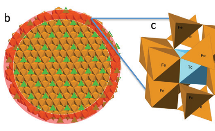
Structural investigation of novel anode materials for solid-oxide fuel cells
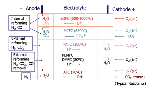 Fuel cells transform the chemical energy produced by oxidation of a fuel into electrical energy with high efficiency and low‐emission of pollutants. The main limitations of current devices are cost and cell lifetime so many studies are underway to develop novel materials for use in the different types of fuel cell.
The Spectroscopy Village is continually developing and upgrading both hardware and software, increasing performance, versatility and reliability of the instruments. B18 updated data acquisition hardware for improved quick Extended X-ray Absorption Fine Structure (EXAFS), I18 implemented a capillary micro reactor for chemical tomography using X-ray diffraction, fluorescence or X-ray Absorption Near Edge Structure (XANES) and I20 increased its versatility in specimen environments. The I14 and I21 beamlines under construction make excellent progress and are expected to be delivered in time and to full performance to the user communities. A major highlight is the progress at I08, which is now open to users with the complete portfolio of imaging and spectromicroscopy modes. I14, I08 and I18 are deeply involved in the Diamond Mapping Project, which is a major revision of the data acquisition software for scanning instruments.
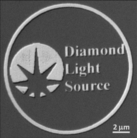
- Figure 1: Differential phase contrast micrograph of a test pattern imaged at I08. Patterns in the letters are, according to the manufacturer (ZonePlates.com), smaller than 20 nm.
I08 received first evaluations of beamline performance from a set of commissioning and user experiments using the entire 250-4400 eV photon energy range. The multi-detector setup with an electron-multiplied CCD camera for detecting transmission signals (absorption and phase-sensitive contrasts) and a large-area silicon drift diode (SDD) for low-energy X-ray Fluorescence (XRF) has been very welcomed. The SDD detector with a very low electronic noise allows detection of elements down to Boron. Typical data acquisition times in the 1ms per pixel range allow fast acquisition of image stacks for Near Edge X-Ray Absorption Fine Structure (NEXAFS) analysis. A number of experiments have successfully been performed freezing radiation-sensitive specimen with cold N2 gas down to temperatures of about -160°C, and keeping such temperatures stable for up to eight hours without observing any degradation of lateral resolutions at the level of 40 nm. In the vicinity of the beamline, I08 installed a dedicated cyro preparation lab for the complete cryogenic workflow from sample freezing to transferring it into the end station.
B18 is improving its already very good capabilities on several fronts. On the detection systems side the beamline has new voltage-to-frequency converters (from the ESRF) which give better digitisation of the ion chamber signals. This leads to an improved S/N when running QEXAFS particularly quickly (< 1min). B18 has also introduced the 36-element Ge fluorescence detector which is giving an improvement over the original 9-element C-train system. While the performance in terms of energy resolution is not as good, the additional fractioning and the increase in collection area give a significant improvement for high count rate experiments in particular. As a result, recent measurement on copper (Cu) adsorbed on heavy matrixes has shown a fourfold flux improvement. On the other hand, where the experiment is photon limited, only the increased solid angle contributes to the improvements. Data on dilute uranium (U) and technetium (Tc) showed improvement by a factor of only two. However, this still represents a significant saving of time and better quality data. On the sample environments side, B18 has commissioned a new hydrothermal cell with pressures up to 40 bar H2. A low temperature system is now available for low energy experiments in a moderate vacuum.
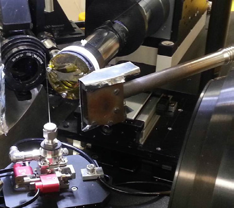
- Figure 2: A capillary micro-reactor mounted on nano-positioning stages on I18 for chemical tomography of single particles. Surrounding the capillary (l-r) are an optical microscope, XRF detector, heater and XRD camera.
On I18, Stephen Price (Diamond) and Andrew Beale (UCL) have led the development of 3D imaging of catalyst particles in either operando or in situ conditions. Stephen has designed a capillary cell which can be adapted for both gas and liquid phase heterogeneous catalysts (Fig. 2). Catalyst particles are suspended on glass wool to trap them firmly in the capillary, which can be heated either by hot air blowers or an infrared lamp source. The output of the capillary can be connected to the input of a mass spectrometer so that the progress of any reaction may be monitored. Imaging catalyst particles on the micron scale using a combination of μ-XRD-CT, μ-XRF/ μ-XANES is very powerful for identifying the structure and distribution of the active state of a supported catalyst particle.
- Figure 3: The I20 team with the first users (UCL) on the Dispersive branch.
Although most of the time and effort invested in the Scanning branch of I20 during the last year has been directed towards solving the instability problems encountered in the performance of the monochromator, there have been developments in other areas. A single chip Medipix detector has been integrated with the X-ray emission spectrometer. The tests performed to date show a significant reduction in the alignment time and an improvement in the data quality. To complement the low temperature sample environment equipment, an in-house infrared mirror furnace for temperatures up to 1000°C has been developed. It allows radiative heating to completely isolate the sample chamber from the heat source, and is specially designed for the collection of data in fluorescence mode. Its modular design allows for future adaptation into a reactor for in situ studies in the field of heterogeneous catalysis.
The main developments made on the Dispersive branch of I20 have been directed at providing the ancillary sample environment that the beamline needs to perform experiments in operando. A gas delivery system has been designed and procured to equip the beamline with the tools necessary to perform fast switching gas experiments. A flow reactor has been designed to cater for the special requirements of the dispersive configuration, and its very versatile design can accommodate different sizes and materials for the reactor tubes. I14 has also completed installation of its front end and a temporary undulator in March 2015 to enable us to start the X-ray commissioning process. The nano-KB (Kirkpatrick-Baez) mirrors and KB mechanics have been designed. The nano-positioning stages have been ordered and assembly and testing of the KB and positioning system will take place over autumn 2015. The XRF detector, a custom design to maximise solid angle within the limited space available, is progressing with delivery expected by the end of 2015. A sample cooling system based around a dry cryogen Gifford-McMahon cooler has been developed and a test chamber has been built to test the sample mounts and cooling assembly. The designs for the novel radiant panel cooling system for the hutches and local thermal enclosures are complete and testing on prototypes have been very promising with stabilities =10mK over long time periods. Options for sample environments are currently being examined and discussions have started on common sample mounts between the nanoprobe and physical science electron microscopes within the new electron microscopy facility.
During 2014, significant progress has been made on the I21 beamline. Most of the internal beamline components have been designed and ordered and will soon be delivered. One of the key beamline components, the plane grating monochromator, has already achieved excellent vibrational stability and will be installed in summer 2015. With on-schedule installation of the internal beamline, I21 will receive its first light in early 2016. With regards to the external building, the design has finished and construction began in September 2014, with its completion planned in 2015. On the end station and the spectrometer side, the I21 team has been working very constructively and has been making good progress. The concept of the sample chamber, including its internal optics and mechanics, the grating assembly, the detector assembly and the whole mechanical structure of the spectrometer arm have developed significantly. Some of the key components of the spectrometer have already been ordered to allow early off-line commissioning. The installation of the whole external beamline shall start from spring 2016.

Figure 4: The new electron microscopy facility and I14 building under construction.
The Core EXAFS beamline B18 covers a wide energy range (2 to 35 keV), which means all elements heavier than phosphorus (P) in the periodic table can be studied. The experiments are run using a continuous energy scanning mechanism (QEXAFS), that allows a very efficient use of beamtime and is ideal for in situ experiments, so that time-dependent phenomena can be studied with a temporal resolution of a few seconds. In addition, as considerable value is added by combining techniques, provision has been made for wide angle X-ray diffraction studies to be incorporated into the beamline architecture. The combination of good flux and of effective detection systems allows collecting data on samples dilute to the mMol region. The instrument offers a variety of sample environments and the flexibility to integrate set-ups designed by the users. Hence, B18 is contributing to research programs across a wide range of scientific disciplines, e.g. solid state physics and materials, catalysis, chemistry, soft matter, surfaces and biomaterials.
I18 is the Microfocus Spectroscopy beamline where X-ray beams as small as 1/40th the width of a human hair are used to probe the chemistry of materials across the whole range of science. Much of the work on the beamline is within the geosciences and biosciences. An example of the crossover between these fields is the research into the biochemistry of fossils by teams led by Professors Phil Manning and Roy Wogelius of the University of Manchester, who have published four papers featuring I18 work investigating both animal and plant fossils in the last year.
The STFC-developed detector read-out electronics have been installed for the silicon drift detectors on I18. This system enables the two detectors to be used at about three times the count rate possible with their previous readout system. This allows the beamline to take better advantage of its high photon flux, and also now enables larger samples to be chemically mapped. The beamline has also upgraded its X-ray diffraction camera with a faster CMOS-based device, once again improving the speed of each measurement. This has proved particularly useful for 3D-imaging experiments, an area where the beamline has progressed considerably in the past year.
2014 has been a very challenging year for the Versatile X-ray Absorption Spectroscopy beamline, I20. The user programme on the Scanning branch had to be suspended when instabilities developed in the performance of the beamline’s four bounce monochromator. Significant efforts have been made to try to resolve the issues and restore the beamline to operational status. This is expected to happen before the end of 2015.
On a more positive note, work to bring the Dispersive branch of I20 into user operation has progressed well, with it seeing its first user experiment in April 2015. The modifications made to the polychromator have allowed the required spectral bandpass to be achieved. The ancillary sample environment and associated timing electronics and control software have been integrated into the beamline to permit the performance of time resolved experiments on microsecond and longer timescales. To take further advantage of the small focal spot delivered by the energy dispersive spectrometer, a diamond anvil cell has been purchased for the beamline to perform experiments at high pressure. To complete the suite of detectors that are required to cover the anticipated experimental programme, a FReLoN camera has been purchased from the European Synchrotron Radiation Facility (ESRF). This detector is particularly well suited to the study of chemical reactions on timescales slower than the millisecond.
I08-SXM is the Scanning X-ray Microscopy (SXM) beamline for morphological, elemental and chemical speciation on a broad range of organic-inorganic interactions in a 250-4400 eV photon energy range, which is unique for an SXM facility. I08-SXM offers versatile imaging modalities such as absorption, phase contrast and dark-field imaging as well as XRF and Near Edge X-Ray Absorption Fine Structure (NEXAFS) spectroscopies. A year ahead of the original project plan, the beamline welcomed first users in July 2014 and performed a series of successful experiments on environmental and earth sciences, material science, bio- and nanotechnology as well as medical applications. Although still in optimisation phase, the beamline already offers the full photon energy range to users with a spatial resolution better than 40 nm for some acquisition modes. A unique capability for a soft X-ray SXM, cryogenic cooling of radiation-sensitive specimen is available as standard for user experiments on the beamline. The I08 team reinforced their effort for establishing a second facility for soft X-ray diffraction imaging (ptychography), pushing the lateral resolution and chemical speciation to new frontiers not achievable with conventional SXM.
I14, the Hard X-ray Nanoprobe beamline, is 185 m long, designed to deliver nanoscale X-ray beams (<50 nm) for elemental, chemical and structural studies of a wide range of materials. Constructing a long beam requires optics hutches within the synchrotron building to condition the X-rays and an experimental endstation housed in an external building to focus the beam and provide the experimental facilities needed to conduct nanoscale studies.
The construction of the external building, which will house the beamline end station, support labs and new electron microscopy facilities, is now well underway with building completion due in September 2015. The I14 team has been busy this year completing the optics hutches and installing the mirrors, monochromator, diagnostics and slits that need to be in place before the X-ray beam is extended to the external building. Over the next year the optics hutch equipment will be extensively tested and commissioned while preparing to build and install the nanoprobe experimental end station. First light is expected in spring 2016 and first users in spring 2017.
The I21 beamline is a dedicated Inelastic X-ray Scattering (IXS) facility at Diamond, focusing on the soft X-ray energy range (250 – 3000 eV) with unprecedented energy resolving power (a few tens of thousands at 1 keV). The beamline will be 81 m long, with its end station and spectrometer accommodated in an external building adjacent to the Diamond ring. By studying the energy and momentum differences between the incident and the outgoing X-rays, users of I21 will be able to investigate the electronic, magnetic and lattice dynamics of novel materials of interest in both condensed matter physics and material science. Systems with strong electronic interactions and new functional materials such as Mott insulators, high-temperature superconductors, oxide heterostructures, catalysts, and carbon-based materials. The beamline construction has made significant progress, with the completion of the internal hutches in June 2014 and the completion of internal cabins and services in March 2015. First light is expected in February 2016 and I21 will welcome its first users in spring 2017.

Diamond Light Source is the UK's national synchrotron science facility, located at the Harwell Science and Innovation Campus in Oxfordshire.
Diamond Light Source Ltd
Diamond House
Harwell Science & Innovation Campus
Didcot
Oxfordshire
OX11 0DE
Copyright © Diamond Light Source. Diamond Light Source® and the Diamond logo are registered trademarks of Diamond Light Source Ltd
Registered in England and Wales at Diamond House, Harwell Science and Innovation Campus, Didcot, Oxfordshire, OX11 0DE, United Kingdom. Company number: 4375679. VAT number: 287 461 957. Economic Operators Registration and Identification (EORI) number: GB287461957003.