Officially opened in 2008, the Membrane Protein Laboratory (MPL) is a research and training facility for scientists interested in solving the 3D structures of membrane proteins by X-ray crystallography. Funded by the Wellcome Trust, the MPL is a joint venture between Diamond and Imperial College London.
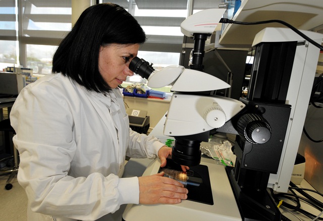
Figure 1: Isabel de Moraes, MPL Coordinator and Group Leader responsible for the management, user projects and developments of the laboratory.
Membrane proteins are important pharmaceutical targets with 60% of the current marketed drugs targeting this class of proteins. However, a major challenge in designing drugs to target membrane proteins is the need for high resolution structural information. With the MPL situated just metres from Diamond’s Microfocus Macromolecular Crystallography (MX) beamline (I24), world-class expertise and tools are brought together to tackle this challenge. The use of Synchrotron Radiation Circular Dichroism (SRCD) through the Circular Dichroism beamline (B23) has also proven to be a valuable high throughput (HTP) screening tool to optimise detergents, ligands and protein batches prior to crystallisation. Furthermore, with the tremendous pace of technological innovation within structural biology, and the co-location of the UK-XFEL hub and the UK National Bio-Imaging Centre (eBIC) at Diamond, the MPL is establishing CryoEM and SFX as viable routes for Diamond-MPL users. The MPL engagement with these methods will definitely have a positive impact on membrane protein structural research.
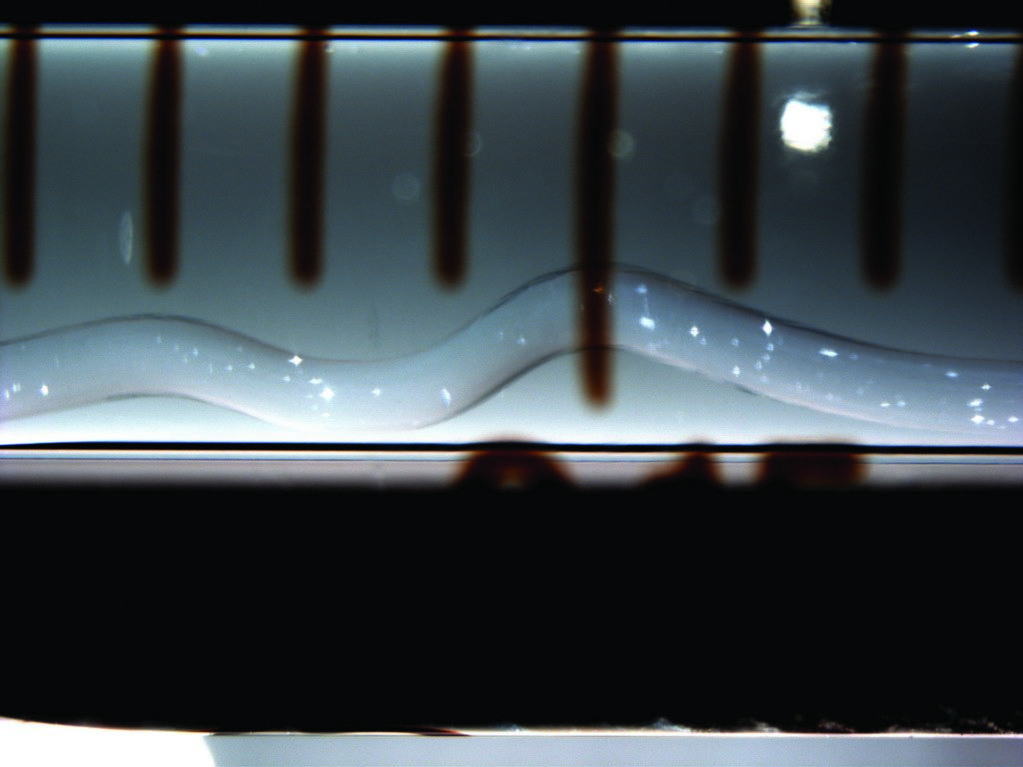
Figure 2: Membrane protein crystal samples prepared for transit to the LCLS.
Since operations began, the MPL has contributed to solving 18 membrane protein structures in total. Following its set up phase from 2007 to 2010, the MPL saw over 210 user visits from 2010 to 2013 during its second phase. The third phase runs from 2013 to 2016 and has already seen almost 200 user visits so far. In 2014, the following were solved with the support of the MPL: the X-ray structure of a CDP-alcohol phosphatidyltransferase; the crystal structures of the human histamine H1 receptor bound to a second-generation and third-generation antihistamines; and the crystal structure of endotoxin modifying enzyme (LptA) from Neisseria meningitides. There are currently around 40 projects in progress, each one usually running over a period of two to three years.
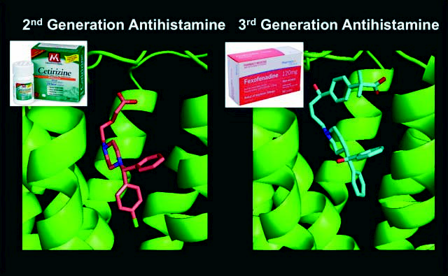
- Figure 3: Structure of the human H1 receptor bound to the highly selective second and third generation antihistamines from X-ray data collected on beamline I24 by the MPL. These new structures will make a significant contribution to computational guided structure-based drug discovery of new antihistamine drugs targeting H2, H3 and H4 receptors, where crystal structures are still absent (manuscript in preparation).
Since 2011, the MPL has been running a hands-on training workshop each year on various aspects of the processes involved such as how to produce a protein, crystallise it and get the best out of it using the Diamond beamlines. Most recently the MPL held a symposium in collaboration with the Industrial Liaison team at Diamond on Membrane Protein Structure Determination for industrial scientists. The event brought together structural biologists and biochemists from a wide selection of pharmaceutical companies, and covered recent approaches devised to address the major challenges related to the expression, extraction, purification and crystallisation of membrane proteins. It also focused on the latest developments in X-ray crystallography experiments at Diamond to obtain structural information from these tiny, extremely fragile, but very important, crystals.
The MPL was invited to take part in a recent G-protein coupled receptor (GPCR) structure, function, drug discovery and crystallography conference at the Royal Society. GPCRs are the targets of over half of all prescribed drugs today (Fig. 3). There is a database of around 800 proteins classified as GPCRs, but drugs have only been developed against 50 of these. The international conference brought together world-leading researchers from industry and academia to discuss recent progress and highlight key areas of future research needed to accelerate GPCR drug-discovery1. One of the key points to emerge is the promise of complementary data gathered from free electron lasers (FELs) as they become more widely available for use. Overall, the conclusion of the meeting was that collaboration between academia and industry is the key to making substantial progress in GPCR drug discovery.
The future looks exciting for the MPL with the laboratory almost fully booked for 2015 and with scheduled beamtime at the FEL in California, USA (LCLS), and the FEL in Hyogo, Japan (SACLA). This will be the first time membrane proteins grown in the UK will be studied using a free electron laser. The MPL has now extended its capabilities to provide support for other scientists who wish to prepare membrane protein samples for FEL experiments (Fig. 2).
References
1.A. Heifetz, G.F.X. Schertler, R. Seifert, C.G. Tate, P.M. Sexton, V.V. Gurevich, D. Fourmy, V. Cherezov, F. H. Marshall, R.I. Storer, I. Moraes, I.G. Tikhonova, C.S. Tautermann, P. Hunt, T. Ceska, S. Hodgson, M.J. Bodkin, S. Singh, R.J. Law and P. C. Biggin. GPCR Structure, Function, Drug Discovery and Crystallography: Report from Academia-Industry International Conference (UK Royal Society) Chicheley Hall, 1-2 September 2014. Naunyn-Schmiedeberg’s Archives of Pharmacology 2015.

Diamond Light Source is the UK's national synchrotron science facility, located at the Harwell Science and Innovation Campus in Oxfordshire.
Diamond Light Source Ltd
Diamond House
Harwell Science & Innovation Campus
Didcot
Oxfordshire
OX11 0DE
Copyright © Diamond Light Source. Diamond Light Source® and the Diamond logo are registered trademarks of Diamond Light Source Ltd
Registered in England and Wales at Diamond House, Harwell Science and Innovation Campus, Didcot, Oxfordshire, OX11 0DE, United Kingdom. Company number: 4375679. VAT number: 287 461 957. Economic Operators Registration and Identification (EORI) number: GB287461957003.