
Beamline Phone Number:
+44 (0) 1235 778709
Principal Beamline Scientist:
Larissa Ishibe-Veiga
Tel: +44 (0) 1235 778869
E-mail: [email protected]
Email: [email protected]
Tel: +44 (0) 1235 778056
Samples must be ultra-high vacuum compatible. The PEEM operates at pressures better than 1x10⁻⁹ mbar and hence any material that degasses is not allowed.
Sample surfaces must be CONDUCTIVE. There is no possibility of imaging insulating surfaces because of charging effects. Ultra-thin oxide layers on conductive substrates can be used. In case of doubt, please ask a member of beamline staff.
The sample is effectively part of the microscope optics: it is biased at the electron acceleration potential of 20KV therefore, to avoid arching towards the grounded objective lens, the sample surface must be SMOOTH. Canted facets or tipped object cannot be focused and may trigger sparks.
The sample is mounted on customized cartridges and is kept in position with a rounded Mo-caps that protect the sample edges from arching (see Figure 1). The outer diameter of the cap is 14 mm and the inner is 10, 7, 4, or 2 mm. The thickness of the sample should not exceed 4 mm.
Samples with a diameter smaller than 4 mm can be mounted using UHV compatible and conductive glue (usually silver paste) or with custom methods to be discussed with beamline staff prior to the experiment.
CAPPING LAYERS can prevent oxidation or make the sample surface conductive, but they should not be thicker than ~2nm. Thicker caps tend to mask the XAS signal excessively, making imaging hard or impossible. As a rule of thumb, aluminium prevents oxidation and is very transparent to low-energy electrons, but it should not be used as an electrode. Metals such as Pt, Ru, and Rh are less transparent to electrons but make good electrodes.
Extra care should be taken when litographic processes are applied to the sample.
Please contact the beamline staff to discuss preparing and mounting your samples well before the experiment.
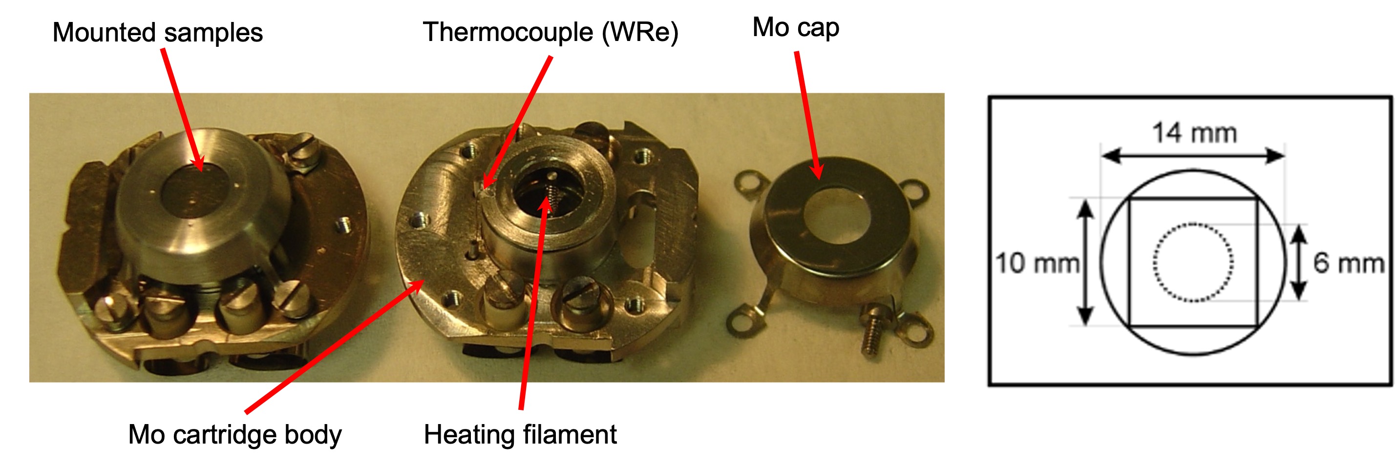
Fig. 1: Left: Elmitec standard cartridge. Right: Schematic drawing of the range of suitable sample sizes.
Samples can be cooled with liquid Nitrogen down to ~ 100 K and can be heated radiatively or with e-beam. Temperatures of approx 1000 K can be reached with radiative heating and 1900 K with e-beam flashing of thin samples. The temperature is measured either with a W/Re thermocouple type C spot-welded on a Ta in thermal contact with the backside of the sample surface or with an optical pyrometer in the preparation chamber.

Diamond Light Source is the UK's national synchrotron science facility, located at the Harwell Science and Innovation Campus in Oxfordshire.
Copyright © 2022 Diamond Light Source
Diamond Light Source Ltd
Diamond House
Harwell Science & Innovation Campus
Didcot
Oxfordshire
OX11 0DE
Diamond Light Source® and the Diamond logo are registered trademarks of Diamond Light Source Ltd
Registered in England and Wales at Diamond House, Harwell Science and Innovation Campus, Didcot, Oxfordshire, OX11 0DE, United Kingdom. Company number: 4375679. VAT number: 287 461 957. Economic Operators Registration and Identification (EORI) number: GB287461957003.

 Magnetic Materials