Instruments by Science Group
I04-1 Contact
Beamline Phone Number:
+44 (0) 1235 778979
Principal Beamline Scientist:
Chris Orr
Tel: +44 (0) 1235 567435
E-mail: [email protected]
Science Group Leader
Dave Hall
Email: [email protected]
Tel: +44 (0) 1235 778926
I04-1 is a fixed wavelength monochromatic beamline which can be found in Zone 05 (orange section) in the Sychrontron Building, located between I03 (anti-clockwise) and I04 (clockwise)
We are optimised for high-throughput data collection with a simple beamline design and streamlined data collection cycle, best suited to well-diffracting crystals, typical of XChem-generated samples.
I04-1 is the primary beamline for data collection of samples from the XChem User Programmes, details can be found at the webpages below:
Data Collection options that we support:
- SPINE pins
- Unipucks
- Fixed wavelength
- XChem
Data Collection options that we do not support:
- Kappa
- Fluorescence
- In situ data collection
- Room Temperature
- Humidity
.png)


WEBCAMS
- Live view! (intranet)Goniometer and Experiment Overview
I04-1 Specification
| Energy | fixed, monochromatic: 0.920Å / 13.53 keV |
| Experimental phasing | SAD: optimal for Br, good for Se |
| Flux (ph/sec) |
3.8 x 10¹² (70um aperture, 300mA ring current) 2.5 x 10¹² (50um aperture, 300mA ring current) 1.3 x 10¹² (30um aperture, 300mA ring current) 9.0 x 10¹¹ (20um aperture, 300mA ring current) 3.0 x 10¹¹ (10um aperture, 300mA ring current) |
|
Beam size options (µm): |
|
| Detector | Eiger 2 XE 9M |
| Maximum resolution (Å) | 1.6 |
| Dataset time, typical | 7.2s (3600 Images, 0.1 degrees, 100% Transmission, 0.002s) |
| Sample changer | Diamond BART with unipucks |
| Sample exchange time | < 20s |
| No. of pins in dewar | 592 |
| Pins and Pucks | SPINE standard pins (only!) and Unipucks |
|
Samples rate (xtals/hour) |
15-30 (manual, user centring) 29 (queued, automatic X-ray centring) |
Please discuss your requirements with a member of the beamline team before your experiment.
SAD phasing at I04-1
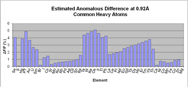
I04-1's wavelength is fixed at the Bromine K edge (E = 13530 eV, λ = 0.9163 Å), where a strong anomalous signal can be measured for most commonly-used heavy atom derivatives (see figure). SAD is usually very effective here - unsurprisingly: thanks to modern detectors and phasing algorithms, tunability is essential only for special cases, especially if the experiment is done carefully (see e.g. Krojer, Pike, von Delft, Acta D 2013)
Estimated anomalous difference at 0.92Å resolution for common elements. These were calculated assuming: for all elements, 1 fully occupied heavy atom site per 300 residues; for Se, 1:42 heavy atoms:residue; and using the formula derived from Smith, J., Curr. Opin. Struct. Biol. vol1, p1002 (1991).
Everything you need to know about shipping samples for data collection on I04-1
Please see the common MX webpages for up-to-date information on sample shipment, here

Diamond Light Source is the UK's national synchrotron science facility, located at the Harwell Science and Innovation Campus in Oxfordshire.
Diamond Light Source Ltd
Diamond House
Harwell Science & Innovation Campus
Didcot
Oxfordshire
OX11 0DE
Copyright © Diamond Light Source. Diamond Light Source® and the Diamond logo are registered trademarks of Diamond Light Source Ltd
Registered in England and Wales at Diamond House, Harwell Science and Innovation Campus, Didcot, Oxfordshire, OX11 0DE, United Kingdom. Company number: 4375679. VAT number: 287 461 957. Economic Operators Registration and Identification (EORI) number: GB287461957003.