____________________________________
Industrial Liaison Group:
Tel: +44 (0) 1235 778797
E-mail: industry@diamond.ac.uk
The World Health Organization estimates that worldwide, annual influenza epidemics result in about 3-5 million cases of severe illness and about 250,000 to 500,000 deaths. Influenza A viruses are the most common cause of seasonal flu in humans and represent a significant risk to public health.
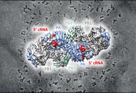
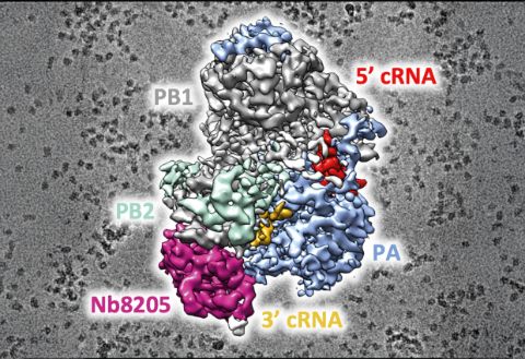
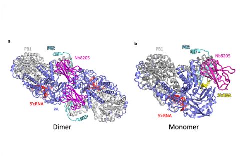
When the influenza virus infects a host cell, it starts to make copies of itself as the disease spreads. The protein driving this behaviour is the viral RNA polymerase which replicates the viral RNA genome and makes RNA templates for protein synthesis. In order to understand how this process works, you need to work out the structure of the RNA polymerase at the atomic level.
Working on the hypothesis that viral replication activates from the formation of the dimer (a form of the protein made of two identical molecules), a team of scientists from the University of Oxford, led by Prof. Jonathan Grimes and Prof. Ervin Fodor, and supported by Wellcome Trust and MRC funding, used a range of techniques at Diamond to determine the structure of the protein.They wanted to find out if they could disrupt the activity of the polymerase by forcing it into a monomeric form.
The team mutated various parts of the dimer interface to produce a selection of mutant proteins that could not form dimers. These results showed that a key step in the replication of the viral genome was blocked if dimerisation was prevented from occurring.
The scientists analysed the structures of the viral polymerase in situ using cryo-Electron Microscopy (cryo-EM) at Diamond’s eBIC facility. They then combined the results with data captured from X-ray crystallography and Biological Small Angle X-ray Scattering (BioSAXS). By studying the live cells, the team was able to see the dimers forming in real-time and be sure that they were necessary for the polymerase to function.
The researchers saw the dimeric form of the polymerase was formed by a specific region of protein and when the region was disrupted, the polymerase couldn’t work. These findings were therefore key in identifying an effective target for new antiviral drugs.
This research was made possible by combining different structural and molecular biology techniques to create a dynamic view of the polymerase. It was greatly facilitated by the unique co-location of Diamond’s cryo-EM facility with other powerful research techniques.
The study has immediate applications for new drug and flu treatments, providing a new effective approach to inhibit the virus in the future.
“The cryo-EM imaging revolution has made a huge impact on our understanding of Influenza virus replication. The location of eBIC at Diamond allows structural biologists, such as myself, to use EM imaging combined with the other X-ray scattering techniques available at the Diamond synchrotron to study proteins like the RNA polymerase of Influenza virus – the part of the virus that drives viral replication.
That these facilities exist in one place and are available to the scientific community is a hugely valuable resource.”Jonathan Grimes, Head of Oxford Particle Imaging Centre, Division of Structural Biology, Wellcome Centre for Human Genetics, Nuffield Department of Medicine, University of Oxford.
Diamond Light Source is the UK's national synchrotron science facility, located at the Harwell Science and Innovation Campus in Oxfordshire.
Copyright © 2022 Diamond Light Source
Diamond Light Source Ltd
Diamond House
Harwell Science & Innovation Campus
Didcot
Oxfordshire
OX11 0DE
Diamond Light Source® and the Diamond logo are registered trademarks of Diamond Light Source Ltd
Registered in England and Wales at Diamond House, Harwell Science and Innovation Campus, Didcot, Oxfordshire, OX11 0DE, United Kingdom. Company number: 4375679. VAT number: 287 461 957. Economic Operators Registration and Identification (EORI) number: GB287461957003.