
___________________________________
Industrial Liaison Group:
Tel: +44 (0) 1235 778797
E-mail: [email protected]
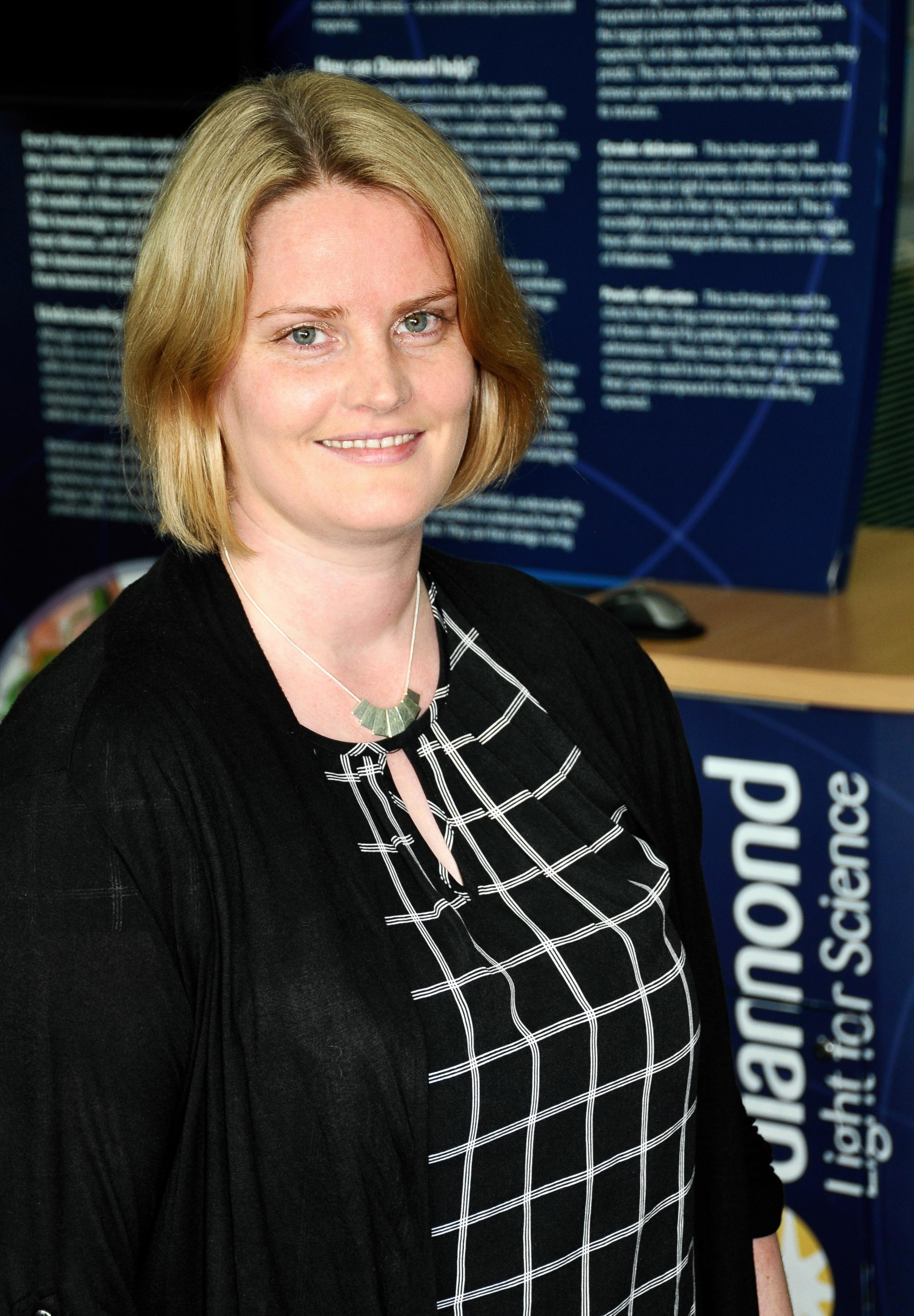
Sally Irvine joined the Industrial Liaison team in October 2015 as an Industrial Liaison Scientist specialising in X-ray imaging. After completing her PhD with the School of Physics at Monash University in Australia in 2010, Sally spent over 3 years as a post-doctoral fellow working within the tomographic microscopy group (TOMCAT) at the Swiss Light Source. Following that role and prior to her position here at Diamond, Sally was a Research Officer for the Laboratory for Dynamic Imaging at Monash University.
With a background specialising in X-ray phase contrast and ultra-fast tomographic imaging, Sally has many years of experience gained from experiments conducted on synchrotron beamlines in Japan, Australia, the US, Switzerland and now the UK. This is well complemented by her research within live-imaging applications of high flux micro-focus laboratory sources.
Sally works closely with industrial users in order to assist them in taking full advantage of the diverse range of imaging capabilities on offer at a selection of Diamond’s beamlines.
Recent advances in instrumentation have led to a dramatic increase in both the speed and resolution of X-ray imaging techniques. Imaging techniques can be applied to a vast range of real world research and development challenges in fields as diverse as pharmaceuticals, automotive engineering, oil recovery and consumer products development.
Hard X-ray imaging allows detailed information to be gathered from below the surface of a material through either full-field imaging, where the whole sample is illuminated, or through scanning, where the beam is focused to a small spot which is scanned across the sample. The high intensity and energy of synchrotron X-rays makes it possible to image a much larger range of materials and sample thicknesses than conventional X-ray sources, and the brilliance of the synchrotron source produces very high resolution images. A technique called X-ray computed tomography can create three dimensional reconstructions of the internal sample volume. This makes it possible to view any cross-section of the virtual image at any angle.
At Diamond, the main microscopy and imaging techniques employed are detailed below:
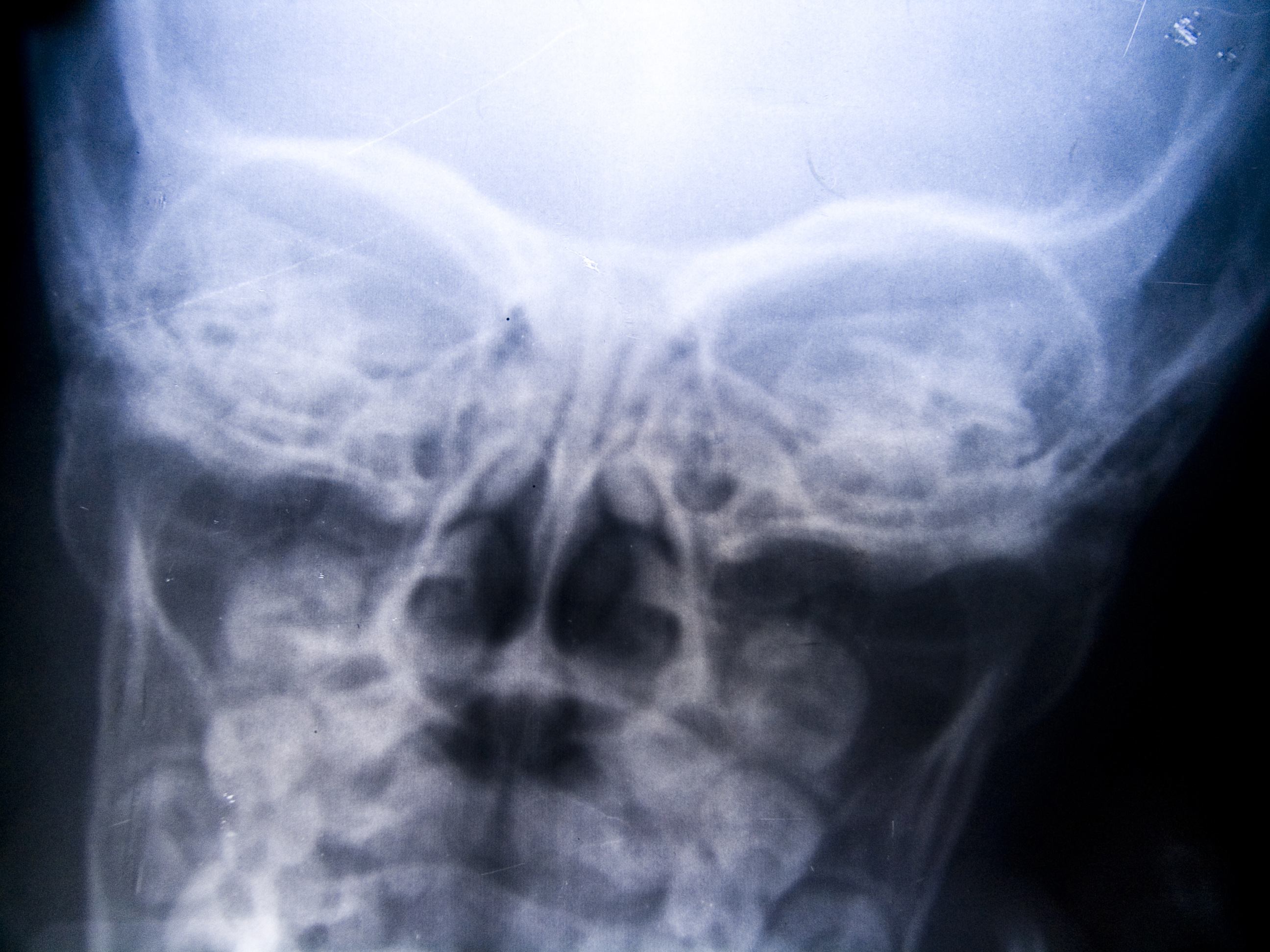
This is the most common imaging technique, and is the technique used in hospital X-ray imaging. An absorption contrast image is essentially a shadowgraph, the contrast being generated by the different attenuating power of materials in the sample. The small spot size and high intensity of synchrotron X-rays also make it possible to scan samples and provide a composite image in much finer detail than from conventional sources. Combining absorption contrast imaging with other X-rays techniques also allows detailed complementary information to be gathered, as in diffraction enhanced imaging, or to recreate three dimensional objects from two-dimensional scans as in tomography.
Phase contrast imaging takes advantage of the fact that different materials have different refractive indices. This produces a phase shift in the X-rays passing through the sample. By placing the imaging detector at a specific distance from the sample, interference between waves can be used to enhance contrast in the image.
It is particularly useful for enhancing the contrast of surfaces and interfaces in samples, which would not be visible using absorption contrast.
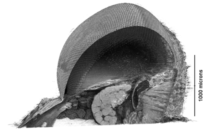
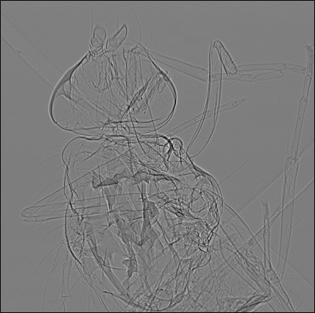
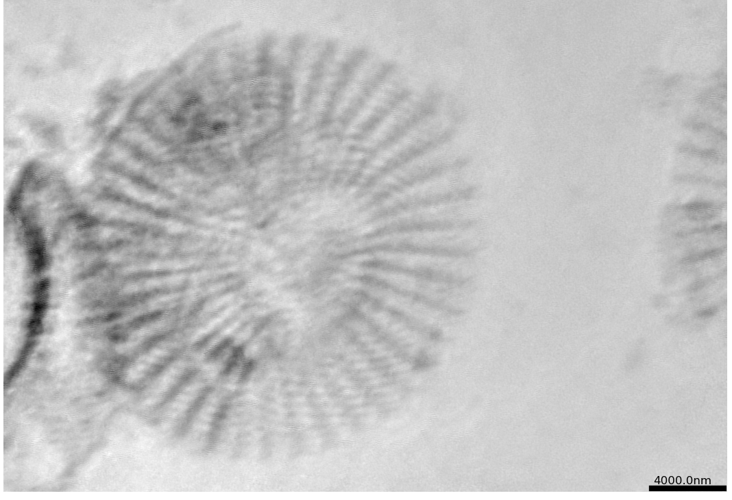
X-ray tomography is the construction of a three dimensional image from two dimensional projections taken at different orientations (usually with phase contrast or absorption contrast imaging). Tomography has many applications in the materials science, engineering and biomedical fields. It can be used to characterise the internal structure of porous materials such as trabecular bone or metal foams. Tomography can be used to determine the size and shape of cracks and other defects inside components such as aircraft parts, where unexpected failures could have catastrophic results. Because it is non-destructive, X-ray tomography can be used to study the internal structure of precious and unique objects in archaeology and palaeontology – for example studying ancient insects fossilised in amber.
Paleontology: Imaging fossilised plankton to understand prehistoric climate change E Read, AJ Bodey & S Redfern.
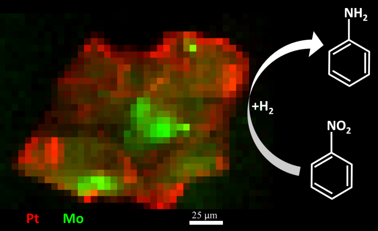
X-ray Fluorescence (XRF) occurs when the inner shell electrons of atoms in the sample get excited by the incident X-ray photons (synchrotron beam) and subsequently release X-ray photon when the system relaxes, that is when the electrons transition from the higher energy levels of the atom to the vacant inner shell. The beauty of this process is that each secondary X-ray photon (sometimes called characteristic radiation) emitted from the sample has a specific energy which is a fingerprint of the atom from which it has originated. By measuring the energy of the secondary photons it is possible to establish the elemental composition of the sample at the point where the X-ray beam hits the sample. Typically a special type of detector called energy-dispersive detector is used to precisely measure the energy of each photon. The plot of the number of photon counts versus their energy, the X-ray spectrum, typically shows a number of peaks which are directly associated with specific elements, so by just glancing at the spectrum it is possible to quickly deduce which elements are present in the sample.
Coherent X-ray diffraction is an imaging technique which overcomes some of the limitations encountered with using lenses. Instead, a series of X-ray diffraction patterns are combined to mathematically reconstruct a three dimensional image of the structure being studied. With highly coherent synchrotron X-rays, this approach can provide spatial resolution on a nm scale. In the past this technique has been applied to model repeating crystal structures, but it is now being used to examine small, non-periodic samples.
The technique is in the very early stages of development and available only to experts through the peer review access route at this stage,

PhotoEmission Electron Microscopy (PEEM) is a non-destructive imaging technique that involves shining linearly or circularly polarised X-rays onto the surface of a sample to provide spectroscopic information on a nm scale. This information can be used to study nanostructures significant for sensors, catalysts, magnetic materials and nanoscale devices and phenomena such as nanomagnetism.
| I12: Joint Engineering, Environmental, and Processing (JEEP) |
 |
| I12 is a high-energy beamline principally for Material Science, Engineering and Processing Science. However, other disciplines are also able to take advantage of the beamline’s high energies and open architecture. The instrument’s main focus is to allow in-situ studies of samples in environments as close as possible to real world environments using imaging, tomography, diffraction and small-angle scattering. I12 is particularly well suited to study large or dense objects and offers a unique sample and environment installation facility for weights up to 2000 kg. |
| Useful for: Chemical imaging, biomedical imaging, engineering and materials science and imaging of components and materials |
| B24: Full field cryo-Xray microscopy |
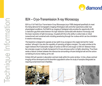 |
| B24 is the Phase III full-field transmission microscope at Diamond designed specifically to meet the rising demand for tomographic imaging of biological specimens under near physiological conditions. The technique bridges the resolution gap that exists between electron microscopy and conventional light microscopy and allows acquisition of tomographic data from both native and fluorescent-labelled samples. |
| Useful for: |
| I06 Nanoscience |
 |
| The Nanoscience beamline exploits the brightest region in Diamond’s spectrum, providing a high photon flux density for soft X-ray experiments. It combines microfocused soft X-rays with variable linear and circular polarisation and X-ray photoelectron emission microscopy (PEEM) to provide spectroscopic data on nanometre length scales. The intense polarised beam can be focused to a spot several microns in diameter, allowing the PEEM to probe nanomagnetism and nanostructures. |
| Useful for: |
| I14 Hard X-ray Nanoprobe |
 |
| Currently under construction, the Hard X-ray nanoprobe I14 beamline will be a dedicated facility for micro-nano SAXS and nanoscale microscopy. The central theme of the beamline is the ability to obtain structural and chemically-specific information on a full range of materials (inorganic/organic) under both static and real (e.g. wet, heated, in-situ strain) conditions. |
| Useful for: Materials science, earth and environmental science and geochemistry, chemistry, biological, biotechnological and biomedical science. |
| I13: X-ray Imaging and Coherence |
 |
| I13 is Diamond’s longest beamline, dedicated to imaging, tomographic and coherence experiments across the biological, medical, geological, material, engineering and archealogical sciences. X-rays are converted to visible light by a scintillator; the visible light is then magnified and collected by a detector. In nano-imaging, an X-ray microscope is used to increase magnification further. A condenser lens known as a Fresnel zone plate focuses the incoming light onto the sample. |
| Useful for: Biomedical research, energy, novel imaging techniques, materials science |
| B22 Infrared Microspectroscopy (MIRIAM) |
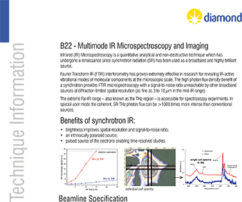 |
| MIRIAM (Multimode InfraRed Microspectroscopy and Imaging) provides a brilliant and versatile microprobe for high resolution imaging of molecular structures as well as high sensitivity vibrational spectroscopy up to the THz range. Experiments span from biomedical applications to physical-chemical researches, with subsequent impact across a wide range of life and physical sciences. |
| Useful for: Surface science, bio-medicine, environmental studies and polymer science |
| I18 X-ray Absorption Spectroscopy |
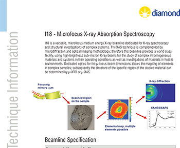 |
| This beamline provides a world class facility, using high-brightness micron-sized X-ray beam for the study of complex inhomogenous materials and systems under realistic conditions. The combination of the brilliance of a third generation synchrotron source, and optics able to focus the beam to a micron sized spot, allows compositional, temporal and spatial information to be gathered at high resolution. On this beamline researchers can map elements in complex samples, follow chemical reactions, study real systems such as mineral samples returned from space, environmental samples and materials in hostile environments. |
| Useful for: |
| I08 Scanning X-ray Microscopy |
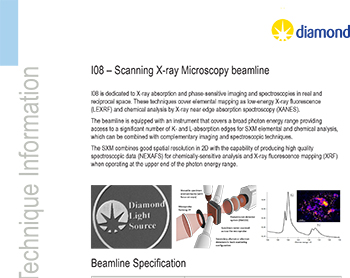 |
| Scanning X-ray Microscopy with variety of imaging and spectomicroscopy modes: Transmission incl. absorption and phase-sensitive contrasts, X-ray fluorescence as well as soft X-ray diffraction imaging (ptychography). |
| Useful for: Earth, environmental science and geochemistry biology and biotechnology, medical and pharmacological science, nanotechnology & material Science |


Diamond Light Source is the UK's national synchrotron science facility, located at the Harwell Science and Innovation Campus in Oxfordshire.
Copyright © 2022 Diamond Light Source
Diamond Light Source Ltd
Diamond House
Harwell Science & Innovation Campus
Didcot
Oxfordshire
OX11 0DE
Diamond Light Source® and the Diamond logo are registered trademarks of Diamond Light Source Ltd
Registered in England and Wales at Diamond House, Harwell Science and Innovation Campus, Didcot, Oxfordshire, OX11 0DE, United Kingdom. Company number: 4375679. VAT number: 287 461 957. Economic Operators Registration and Identification (EORI) number: GB287461957003.

 Industrial Liaison Office