
___________________________________
Industrial Liaison Group:
Tel: +44 (0) 1235 778797
E-mail: [email protected]
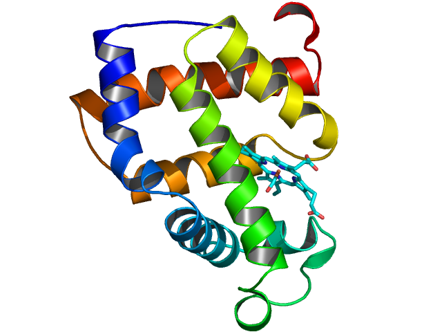
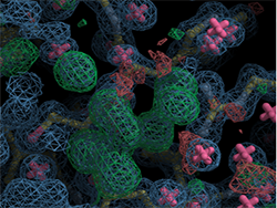
Macromolecular Crystallography (MX, also referred to as Protein Crystallography or PX) is the most powerful method for determining the atomic three dimensional structures of large biological molecules. It is a vital tool for linking structure with function, for rational drug design, for investigating protein folding and for relating other structural information, such as evolutionary relationships, from biological molecules. Diamond is a world leading facility for structural biology. The outstanding design of the macromolecular crystallography (MX) beamlines combines the provision of cutting edge technical instruments essential for de novo structure solution and the study of protein-ligand interactions with the latest developments in automation required to accelerate lead identification and ligand screening processes.
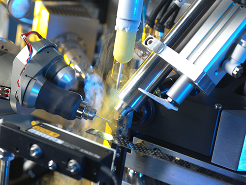
Macromolecules tend to form small, imperfect and weakly diffracting crystals. The high brightness of synchrotron X-rays makes the collection of precise measurements possible. Our state of the art macromolecular crystallography (MX) beamlines are fitted with advanced robotic systems and software for automated sample handling, crystal centering and data collection. Many of the macromolecular crystallography (MX) beamlines conduct in situ experiments which are advantageous as they can be carried out without any manipulation of individual crystals, thus preserving the crystal integrity. In addition, microfocus beamlines are perfectly suited for studying small or disordered crystals and facilities for containment level 3 samples are in place.
At Diamond, we have developed high performance automated processing pipelines for integration, scaling and phasing data in real time for immediate structure analysis directly at the beamline. We follow a programme of continuous upgrades to maintain our world leading position for structural biology research. Beamline specifications are updated frequently as upgrades are introduced onto the beamlines.
Applications
⋅ Rational drug design
⋅ Enzyme mechanisms
⋅ Supramolecular structure
⋅ Molecular recognition
⋅ Nucleic acids
⋅ Structural genomics
⋅ High throughput crystallography
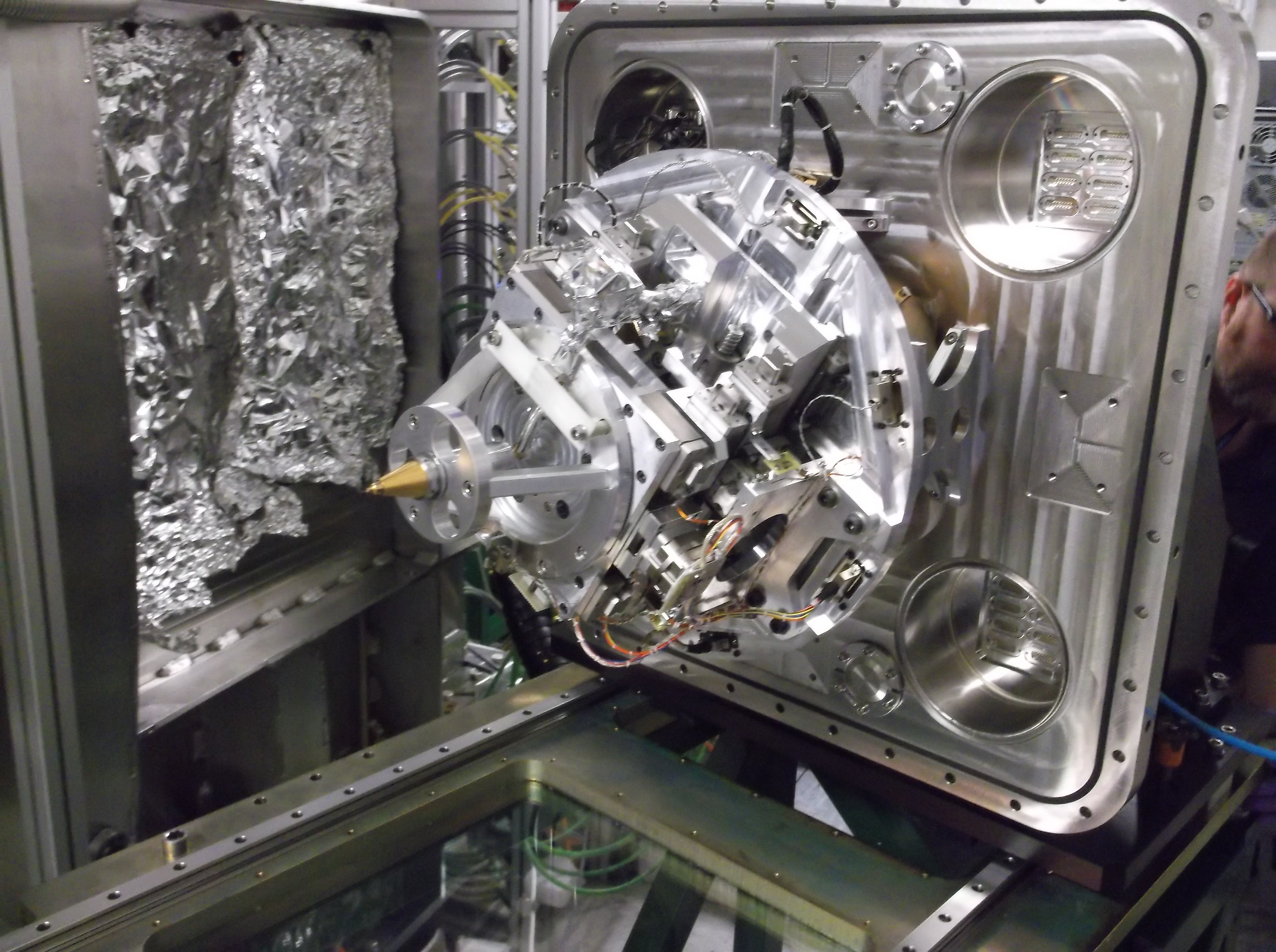
There are currently 5 operational beamlines in the macromolecular crystallography (MX) suite; I02, I03, I04, I04-1 and I24 with 3 further beamlines in development; VMXm, VMXi and I23
I02 and I03 are tuneable beamlines with a working wavelength range of 0.68 - 2.48 Å. The standard working energy is 12.658 keV which provides a wavelength of 0.979 Å and a focussed beam size of 80 x 20 um (FWHM). As with all the MX beamlines, I02 and I03 use SPINE standard pins and Unipucks. I03 also offers in situ experiments using SBS format plates. Additionally, I03 can provide containment measures for experiments involving biological agents in Hazard Groups 2 and 3.

I04 has a typical working wavelength is 0.9795 Å (12.658 keV) but it is tuneable over the wavelength range 0.708 - 2.066 Å. The beamsize can be focussed from 10 x 5 to 110 x 100 microns across the available energy range. As with all macromolecular crystallography (MX) beamlines, I04 uses SPINE standard pins and Unipucks from cryocooled samples. The beamline can be accessed remotely.
I04-1 is a fixed wavelength monochromatic beamline, with its undulator in I04's straight section. The beamline was originally aimed at high-throughput data collection for well-diffracting crystals, with off-the-shelf robotics and a stable beam thanks to a simple beamline design. However, it is now fully part of the routine macromolecular crystallography (MX) user program, for both academic and industrial users alike, since the constraints (5-fold weaker beam than I02/3/4, fixed energy) affect only a subset of macromolecular crystallography (MX) experiments. In addition, the beamline also offers a world-first, namely routine medium-throughtput fragment screening by crystal structure, including all steps from crystal soaking and harvesting to data.
I24 is a tuneable microfocus beamline for macromolecular crystallography (MX) . It was the first beamline of it’s kind to be built in Europe and has been operating since 2008. It is currently in the process of a complete rebuild. The rebuild of the endstation is complete (hardware-wise) and new mirrors are now at Diamond and will be installed early 2016. When complete it will be able to offer a ~2.5x5 micron beam with a state-of-the-art endstation.
The beamline offers extremely high flux densities with the ability to investigate virus structure, membrane proteins, microcrystals (crystals as small as 1.5 microns have been measured successfully). It combines versatile optics and the most sophisticated detectors available with the most advanced automation systems to enable sample location, data collection and analysis.
We also have 3 macromolecular crystallography (MX) beamlines under construction:
VMXm is a micro/nanofocus Macromolecular Crystallography beamline aimed at atomic structure determination in cases where the production of significant quantities of protein material and crystals is problematic. Indeed this is the case for many challenging protein complexes and medically important macromolecules that yield only very small crystals. The X-ray beam size on VMXm will be less than 0.5 microns and with the use of novel X-ray optics and electron beam imaging methods the tiniest protein crystals will be able to be precisely aligned into this X-ray beam, in vacuo, and the resulting X-ray diffraction data measured.
The ability to tune the X-ray energy allows additional information to be obtained from heavy atoms within the macromolecules to aid in structure determination by multicrystal SAD or MAD methods. In many ways VMXm will be a hybrid X-ray/cryoEM instrument making use of methods for sample preparation from cryo-electron-microscopy, imaging from scanning electron microscopy and diffraction data collection methods from X-ray crystallography. VMXm is currently under construction and is schedule for first user operations towards the end of 2017.
The Versatile Macromolecular Crystallography in-situ (VMXi) beamline will be an entirely automated facility for characterisation of, and data collection directly from, crystallisation experiments in situ.
I23 will be a long-wavelength Macromolecular Crystallography (MX) beamline, and will be a unique facility for solving the crystallographic phase problem, using the small anomalous signals from sulphur or phosphorous which are present in native protein or RNA/DNA crystals. This will be of increased importance for projects where protein labelling to introduce anomalous scatterers is not feasible. In addition, the beamline's wavelength range will provide access to the M-edges of elements, with huge anomalous signals offering new opportunities for phasing large molecular complexes.
I23 will complement the existing suite of five macromolecular crystallography (MX) beamlines at Diamond and will be optimised for operation in the wavelength range from 1.5 to 4 Å. The experimental end station will be in vacuum to minimize absorption and scattering effects. A large semi-cylindrical detector will allow measurements of a large range of diffraction angles and a multi-axis goniometer will be available for crystal alignment and orientation. An X-ray tomography setup will be integrated into the beamline end station to obtain the crystal shape and volume as a basis of an analytical absorption correction. First successful data has been collected demonstrating the advantages of in-vacuum macromolecular crystallography. The beamline is currently in commissioning mode welcoming first users in Dec 2015.
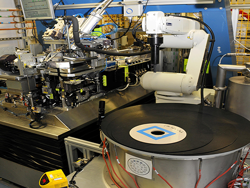
Macromolecular Crystallography (MX) Sample Changer Upgrade (BART)
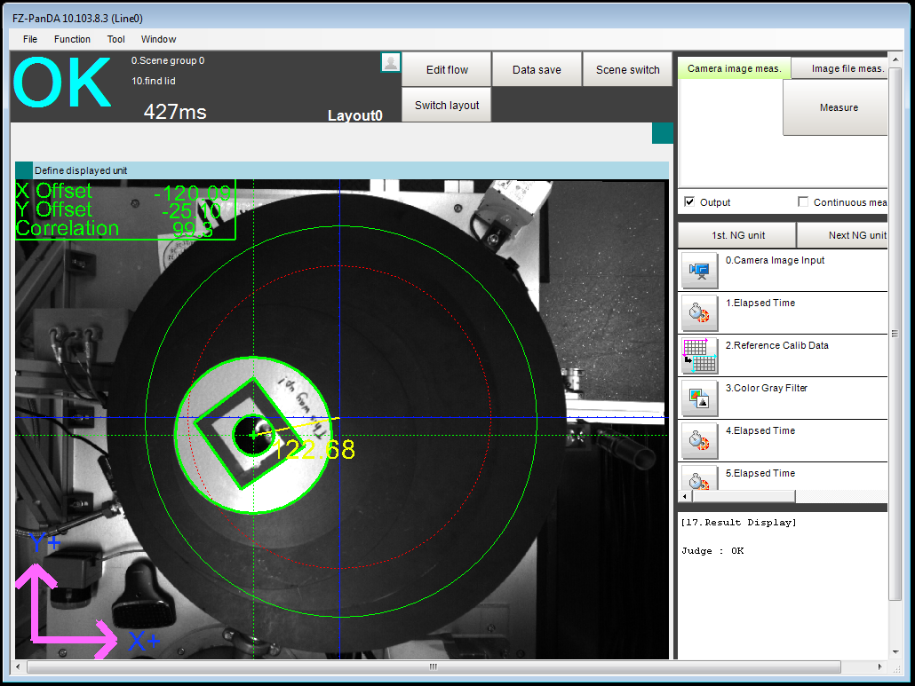 Since January 2015, a new robotic sample changer has been operational on beamline I03. The new robot, BART, has improved exchange times, increased sample capacity significantly and is more tightly integrated into the beamline control systems which means reliability has also improved. Find out more here...
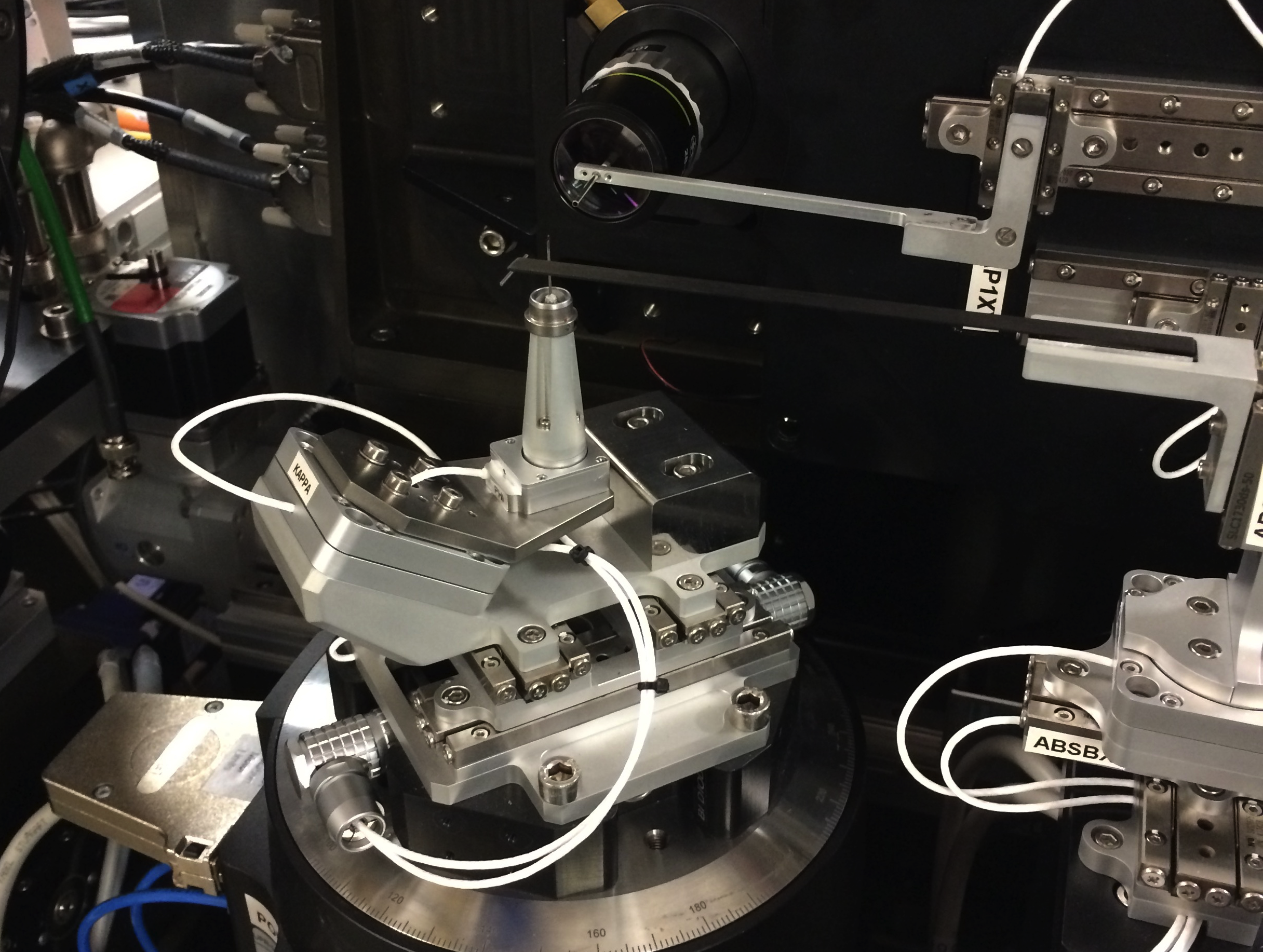
 Recent upgrades to beamline I04 include the implementation of a mini-kappa goniometer, a new focussing system using compound refractive lenses and restructuring of the data collection scripts used by GDA. Read more here....
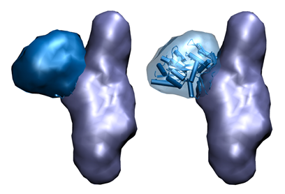
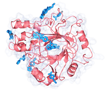 Fragment screening is a well-established approach in the development of novel compounds for drug discovery, yet nowhere has it been accessible as public facility. Diamond are now offering a world-first service called XChem, one-stop-shop that allows screening fragments by the most sensitive technique available, namely X-ray crystallography. Read more here....
 Auto-processing pipelines are now an essential and constantly developing part of the macromolecular crystallography (MX) beamline package. Autoprocessing helps users decide how to collect the best data and plan the most efficient use of the available beamtime. As a result, users not only get the optimum results from their samples but can also often achieve these in shorter timescales. Read more here...
 Solution small angle X-ray scattering (also known as protein SAXS or bioSAXS is a powerful tool for analysis of the structure of proteins in solution. It can be used for a wide range of protein types and solution conditions. Read more here...
• The autoprocessing pipeline xia2 is now able to use the new data processing software DIALS (http://dials.diamond.ac.uk/) for indexing and integration of the diffraction images. In addition, the resolution limit of data automatically processed by xia2 is now determined using CC½ rather than I/sig(I) or Rmerge.
• A New Pilatus 6M detector has been installed on I04-1.
• Automatic alignment strategy for the use of mini-kappa is incorporated in the strategy calculation and easy to find on ISPyB.
• All macromolecular crystallography (MX) beamlines now have an interactive screen in the experimental hutch which allows users to register puck positions in the sample changer dewar. Information about the puck positions is automatically linked to ISPyB and GDA.
• I03, I04 and I04-1 are now fitted with the new sample changer BART which replaced the Actor robots.
MX BAG training takes place 2-3 times annually at Diamond. If you or a colleague would like to take part please let us know on [email protected].
If you wish to talk to the Industrial Liaison Team about your research requirements, then please contact us on +44 (0)1235 778797 or [email protected] or you can follow us on Twitter @DiamondILO or connect with us on Linkedin.

Diamond Light Source is the UK's national synchrotron science facility, located at the Harwell Science and Innovation Campus in Oxfordshire.
Copyright © 2022 Diamond Light Source
Diamond Light Source Ltd
Diamond House
Harwell Science & Innovation Campus
Didcot
Oxfordshire
OX11 0DE
Diamond Light Source® and the Diamond logo are registered trademarks of Diamond Light Source Ltd
Registered in England and Wales at Diamond House, Harwell Science and Innovation Campus, Didcot, Oxfordshire, OX11 0DE, United Kingdom. Company number: 4375679. VAT number: 287 461 957. Economic Operators Registration and Identification (EORI) number: GB287461957003.

 Industrial Liaison Office