
Keep up to date with the latest research and developments from Diamond. Sign up for news on our scientific output, facility updates and plans for the future.
The Optics and Metrology group continue to provide expert support to Diamond’s beamlines in the design, testing, and optimisation of beamline optics. We have also increased the technical capabilities of many existing and future beamlines by pursuing a broad range of optical research projects1-11. Analogous to using contact lenses to correct our eyesight, custom-designed refractive phase correctors were designed to compensate aberrations of non-perfect X-ray mirrors. These novel optics resulted in a ten times improvement in the performance of a prototype X-ray mirror. Ultra-high accuracy characterisation of state-of-the-art X-ray optics continues to be an essential procedure, both for quality control and the development of novel optics. Continuous improvements of metrology instrumentation and techniques has led to ever more accurate at-
 Figure 1: Optical microscope image (in negative) of the refractive structure used as an X-ray wavefront corrector.
Focusing X-rays to nanometre sized focal spots requires high precision X-ray optics. For micro- and nano-focusing, height errors on the mirrors can significantly distort the X-ray beam. This causes blurring of the X-ray focal spot, resulting in a limit to the spatial resolution. We have implemented a method for correcting the reflected wavefront of a focusing mirror by using custom-designed refracting structures upstream of the mirror1. This novel concept was tested using a previously developed prototype multilane mirror for rapidly switching focused beam sizes6. The mirror has three parallel lanes running the full length of the 90mm long reflecting surface, with lane 1 having the basic elliptical shape providing a beam size of ~0.5 μm. Lanes 2 and 3 were deliberately profiled to provide beam sizes of 2 μm and 10 μm respectively. Wavefront distortions from the refractive structures were purposefully designed to cancel out distortions from the mirror. To determine the shape of the refractive structures, surface profile measurements of the mirror were made, with sub-nanometre scale accuracy, using the Diamond-NOM and at-wavelength metrology. Refractive structures (Fig. 1) were fabricated using lithography (LIGA) at Indus-2, India. X-ray measurements on Diamond’s Test beamline (B16) of the hybrid optic wavefront corrector (refractive corrector plus reflective focusing optics) showed a reduction in the size of the focal spot of a multi-lane mirror by a factor more than 10 (Fig. 2). This is equivalent to the effective slope error reducing from ~ 5 μrad to ~ 0.2 μrad rms. This important work, which could have major impacts for Diamond’s X-ray optics, is now being extended and applied to nano-focusing mirrors and refractive X-ray lenses.
APPLE II undulators are used on several beamlines at Diamond to provide high brilliance soft X-ray beams with the capability to switch between various photon beam polarisation states. Accurate knowledge of the polarisation state of the photon beam is crucial to be confident in beamline results. Hence, it is essential to perform in situ polarisation measurements to understand the undulator and beamline performance. Diamond has developed a high-precision soft X‑ray Polarimeter, which utilizes two rotating multilayer optics (retarder and analyzer). The Polarimeter has been used to characterise the polarisation characteristics of Diamond’s soft X-ray beamlines. Recently2, it was employed to systematically investigate the polarisation characteristics of the two helical APPLE II undulators installed on the Nanoscience beamline (I06) at Diamond (Fig. 3).


Figure 2. Focused X-ray beam profile at 10 keV for lanes 2 (left) and 3 (right) before (black curves) and after (blue curves) adding the refractive compensator. Also shown for reference in red is the measured focused beam profile of the unmodified mirror lane 1. Data have been scaled so that each profile has similar peak height.
For each polarisation measurement, data were acquired for a complete rotation of both the retarder and analyzer to provide a map of the detected light intensity which is characteristic of the photon polarisation state. Fig. 4 demonstrates the agreement with theoretical calculations for left-handed circular polarised light. The only discrepancy is a variation in observed peak intensity which indicates the presence of a linear component in the otherwise circularly polarised photon beam. The measurements have brought out the effect of the phasing unit on the polarisation state when the two undulators are phased together for increased flux. Importantly, a negative correlation between the degree of circular polarisation and the photon flux was found when the phasing unit was used. Such precise polarisation measurement provides valuable inputs for any synchrotron facility where dual undulators are used for polarisation-dependent measurements.
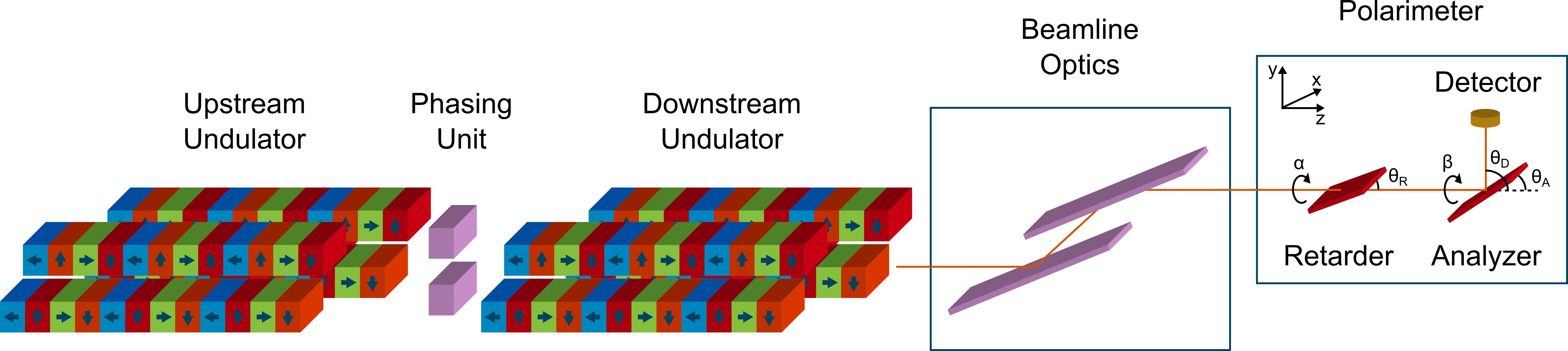
Figure 3. Schematic of the undulator configuration at beamline I06 with polarimeter.
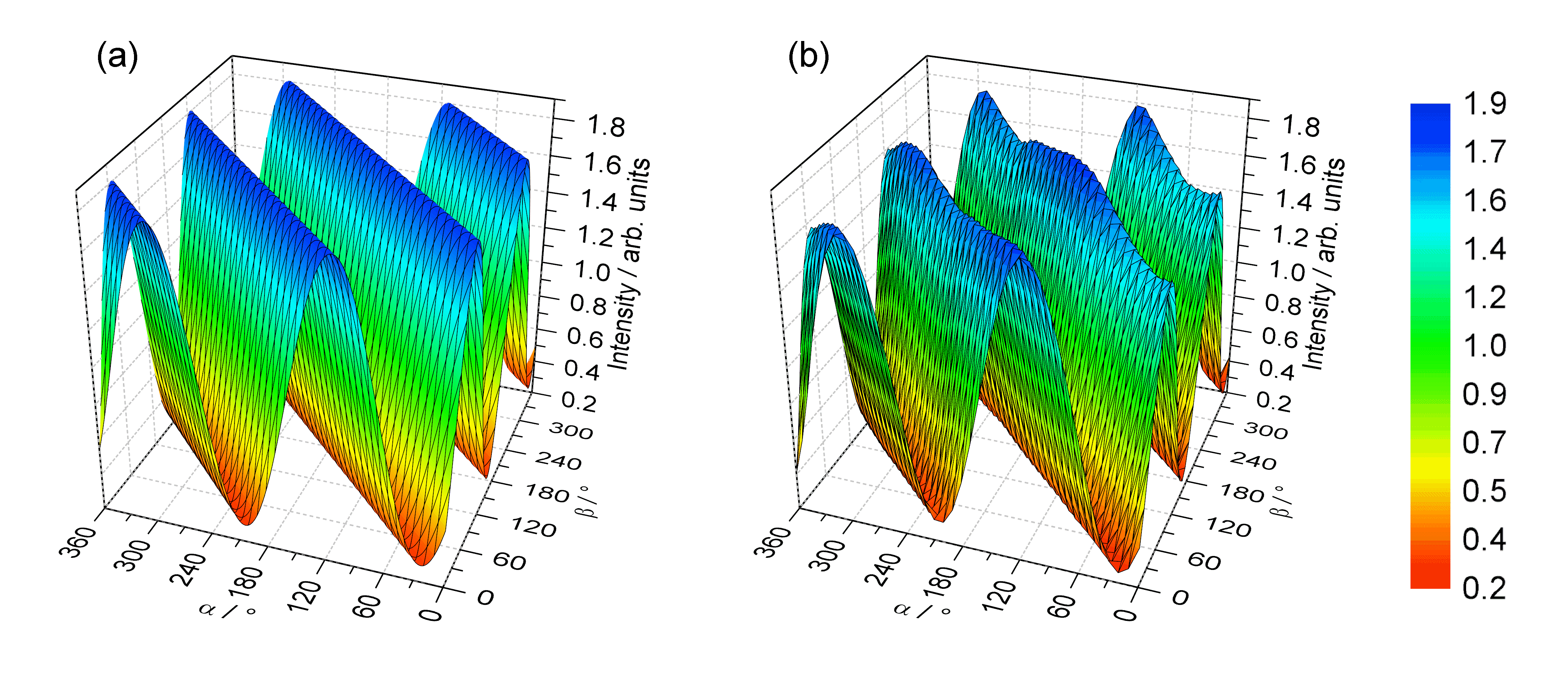
Figure 4. 3D map of left circular polarised light intensity at 375 eV from (a) theoretical calculation, and (b) measurements using the Diamond Polarimeter.
1. Sawhney K., Laundy D., Dhamgaye V., Pape I. Compensation of X-ray mirror shape-errors using refractive optics. Appl. Phys. Lett., 109, 051904 (2016).
2. Hand M., Wang H., Dhesi S.S., Sawhney K. Investigation of the polarization state of dual APPLE-II undulators. J. Synchrotron Rad., 23, 176-181 (2016).
3. Alianelli L., Laundy D., Alcock S., Sutter J.P., Sawhney K. Development of Hard X-ray Focusing Optics at Diamond Light Source. Synchrotron Radiation News, 29, 3-9 (2016).
4. Wang H., Kashyap Y., Cai B., Sawhney K. High energy X-ray phase and dark-field imaging using a random absorption mask. Scientific Reports, 6, 30581 (2016).
5. Sutter J.P., Alcock S.G., Kashyap Y., Nistea I., Wang H., Sawhney K. Creating flat-top X-ray beams by applying surface profiles of alternating curvature to deformable piezo bimorph mirrors. J Synchrotron Rad., 23, 1333-1347 (2016).
6. Laundy D, Sawhney K., Nistea I., Alcock S.G., Pape I., Sutter J., Alianelli L., Evans G. Development of a multi-lane X-ray mirror providing variable beam sizes. Rev. Sci. Instrum., 87, 051802 (2016).
7. Alcock S.G., Nistea I., Sawhney K. Nano-metrology: The art of measuring X-ray mirrors with slope errors <100 nrad. Rev. Sci. Instrum., 87, 051902 (2016).
8. Kashyap Y., Wang H., Sawhney K. Development of a speckle-based portable device for in situ metrology of synchrotron X-ray mirrors. J. Synchrotron Rad., 23, 1131-1136 (2016).
9. Sutter J.P., Boada R., Bowron D.T., Stepanov S.A., Diaz-Moreno S. Rotation of X-ray polarization in the glitches of a silicon crystal monochromator. J. Appl. Crystallogr., 49, 1209-1222 (2016).
10. Kashyap Y., Wang H., Sawhney K. Experimental comparison between speckle and grating-based imaging technique using synchrotron radiation X-rays. Opt. Express, 24, 18664-18673 (2016).
11. Kashyap Y., Wang H., Sawhney K. Speckle-based at-wavelength metrology of X-ray mirrors with super accuracy. Rev. Sci. Instrum., 87, 052001 (2016).

Diamond Light Source is the UK's national synchrotron science facility, located at the Harwell Science and Innovation Campus in Oxfordshire.
Copyright © 2022 Diamond Light Source
Diamond Light Source Ltd
Diamond House
Harwell Science & Innovation Campus
Didcot
Oxfordshire
OX11 0DE
Diamond Light Source® and the Diamond logo are registered trademarks of Diamond Light Source Ltd
Registered in England and Wales at Diamond House, Harwell Science and Innovation Campus, Didcot, Oxfordshire, OX11 0DE, United Kingdom. Company number: 4375679. VAT number: 287 461 957. Economic Operators Registration and Identification (EORI) number: GB287461957003.

 A brighter light for science