Many patients suffering from chronic conditions such as diabetes, schizophrenia and acromegaly are required to administer treatments subcutaneously or intramuscularly using injections.
Whilst the efficacy of these treatments has been well documented, the absorption mechanisms, the impact of different formulations and their delivery, is not yet fully understood.

Investigating novel drug delivery platforms
Case Study

Understanding the journey of the active pharmaceutical ingredients
The challenge
It is critically important to fully understand the route that injected pharmaceuticals take when being absorbed into the body, so that a sustained release of therapeutic agents can be achieved whilst improving patient adherence and tolerability. To do so requires a fast, high resolution method to track the journey, as it happens, across a large sample of tissue.
The solution
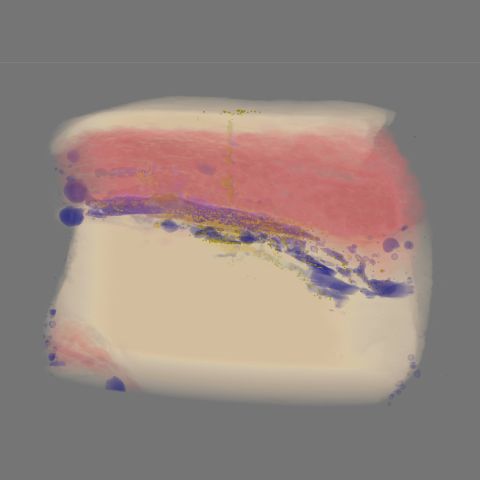
To understand the journey of the active pharmaceutical ingredients (API) within the muscular tissue, scientists from AstraZeneca and Diamond performed high energy X-ray computed tomography, using beamline I12 at Diamond. This instrument provides a sufficiently large X-ray beam to image an appropriate volume of tissue and high intensity X-rays to image the tissue rapidly before it dries out. For this study scientists used active pharmaceutical ingredients encapsulated within biodegradable polylactide-co-glycolide (PLG) microspheres as a drug delivery platform. Samples of porcine tissue were injected with different delivery media all of which contained microspheres (20-100 µm) encapsulating the API.
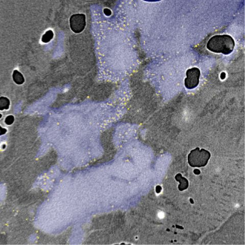
The resulting reconstruction of large volumes of pork tissue allowed the whole picture of the intramuscularly injections to be seen and segmented. The team were able to understand the overall dispersion of the injection media as well as observing the individual microspheres and how they accumulated around the injection site.
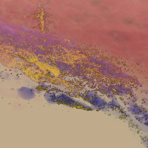
Using micro-tomography at Diamond, scientists were able to rapidly obtain high resolution visualisations of individual microsphere particles and the delivery vehicle media after injection in soft tissue. This research approach enabled scientists to follow the journey of the injected formulations. By doing this, they hope to understand the interplay between patient physiology, formulation and physicochemical attributes. The findings of these investigations will be used to drive the design and development of novel formulations and devices, to provide new effective drug therapies.
Claire Patterson, Dean Murphy, Sarah Irvine, Leigh Connor & Zahra Rattray
Novel application of synchrotron x-ray computed tomography for ex-vivo imaging of subcutaneously injected polymeric microsphere suspension formulations.
Pharm Res 37, 97 (2020). https://doi.org/10.1007/s11095-020-02825-9