
Across the world, billions of people suffer from enamel caries, commonly known as tooth decay. Diets rich in sugar and poor dental hygiene enable bacteria in the mouth to form biofilms (plaque). As they break down sugars and other food debris, the bacteria produce acids that attack the enamel and cause demineralisation. Enamel caries can cause sufferers considerable pain and distress, and remedial treatment is expensive. At present, there is no reliable way of reversing the damage and remineralising enamel. Enamel is a complex mix of mineral hydroxyapatite, water and binding proteins, arranged in a hierarchical structure. Developing effective treatments to prevent and reverse damage to the enamel requires a thorough understanding of how demineralisation proceeds at different hierarchical levels. In work recently published in Materials & Design, a team of researchers from the University of Oxford and the University of Birmingham led by Professor Alexander M. Korsunsky combined wide-angle X-ray scattering (WAXS) with other imaging and analysis techniques to reveal the details of enamel demineralisation across the hierarchical structure levels.
Professor Alexander M. Korsunsky, from the University of Oxford, said:
"This work extends our knowledge of the changes occurring at the sub-micron scale and highlights the importance of using high-resolution X-ray analysis to understand both the underlying roots of disease, and the routes to its treatment."
Tooth decay is a relatively slow process, and so it is challenging to study its progress in real time. In their series of studies, the team artificially demineralised sections of human enamel using lactic acid and compared them to healthy samples using optical and scanning electron microscopy and scanning X-ray diffraction.
Supporting studies using energy-dispersive X-ray spectroscopy (EDS) showed a decrease in the calcium to phosphorus ratio in the etched samples, highlighting the role of preferential removal of calcium.
Optical imaging at the mm scale demonstrated that acid demineralisation of the enamel reduced its birefringence and can help identify demineralised enamel. (The refractive index of birefringent materials depends on the polarization and propagation direction of light.)
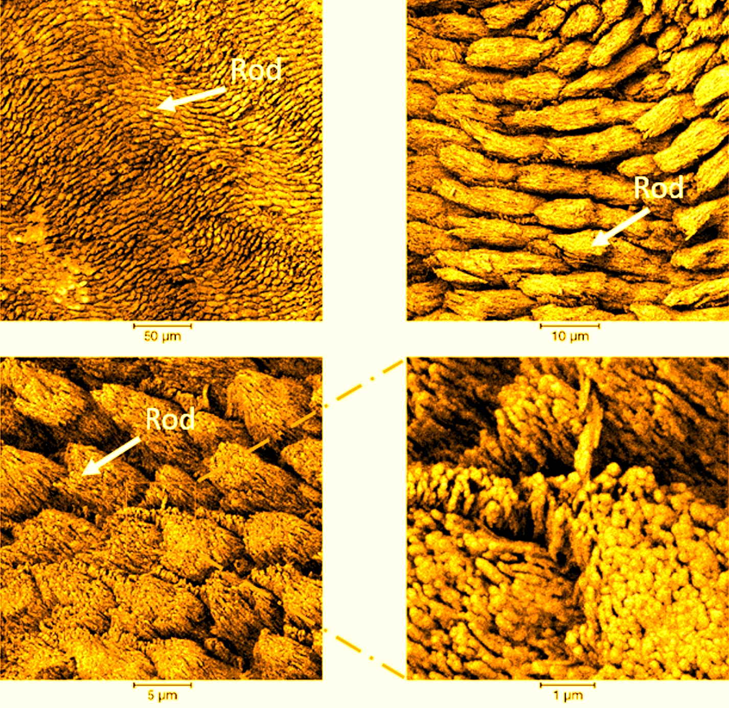
Focused Ion Beam - Scanning Electron Microscopy (FIB-SEM) revealed the structural differences between intact and demineralised samples down to the nanoscale, and confirmed the correlation between the apparent loss of birefringence and nanoscale ultrastructure changes.
At Diamond, the researchers used wide-angle X-ray scattering (WAXS), performed on beamline B16. This allowed them to reveal the differences in the crystal structure of the enamel before and after etching down to the length scale of Ångstroms. They assembled texture maps at 500 nm spatial resolution and correlated them with the enamel structure. This multi-scale correlative approach allowed them to link the structural appearance of the enamel under electron microscope with intricate changes at the level of its crystal lattice.
Lead author Dr Cyril Besnard, from the University of Oxford, explains:
Wide angle X-ray scattering using tightly focused beams is an extremely powerful technique. The high-resolution setup available at beamline B16 allowed us to establish correlated understanding of the structure of enamel spanning seven orders of magnitude, from the macroscale (millimetres) down to fractions of Å. Combining WAXS with other imaging techniques was crucial to provide us with new insights into the acid demineralisation process. Diamond beamline staff were an amazing resource throughout, supporting our efforts at every stage of the experiment.
This research is part of a larger project funded by EPSRC devoted to tackling human dental caries by multi-modal correlative microscopy and multi-physics modelling. The team are continuing their work using the techniques they developed in their labs and at Diamond to combine real and reciprocal space imaging techniques (diffraction and tomography) at Diamond using the new K11 Dual Imaging And Diffraction (DIAD) instrument, as well as I13 Imaging and Coherence and I14 X-ray Nanoprobe beamlines.
To find out more about the B16 beamline or discuss potential applications, please contact Principal Beamline Scientist, Kawal Sawhney: [email protected].
Besnard C et al. Analysis of in vitro demineralised human enamel using multi-scale correlative optical and scanning electron microscopy, and high-resolution synchrotron wide-angle X-ray scattering. Materials & Design 206 (2021): 109739. DOI:10.1016/j.matdes.2021.109739.
Besnard C et al. 3D analysis of enamel demineralisation in human dental caries using high-resolution, large field of view synchrotron X-ray micro-computed tomography. Materials Today Communications (2021): 102418. DOI:10.1016/j.mtcomm.2021.102418.

Diamond Light Source is the UK's national synchrotron science facility, located at the Harwell Science and Innovation Campus in Oxfordshire.
Copyright © 2022 Diamond Light Source
Diamond Light Source Ltd
Diamond House
Harwell Science & Innovation Campus
Didcot
Oxfordshire
OX11 0DE
Diamond Light Source® and the Diamond logo are registered trademarks of Diamond Light Source Ltd
Registered in England and Wales at Diamond House, Harwell Science and Innovation Campus, Didcot, Oxfordshire, OX11 0DE, United Kingdom. Company number: 4375679. VAT number: 287 461 957. Economic Operators Registration and Identification (EORI) number: GB287461957003.

 Science