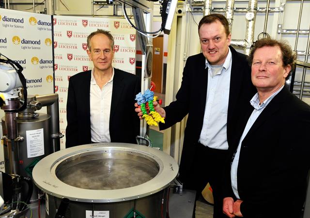
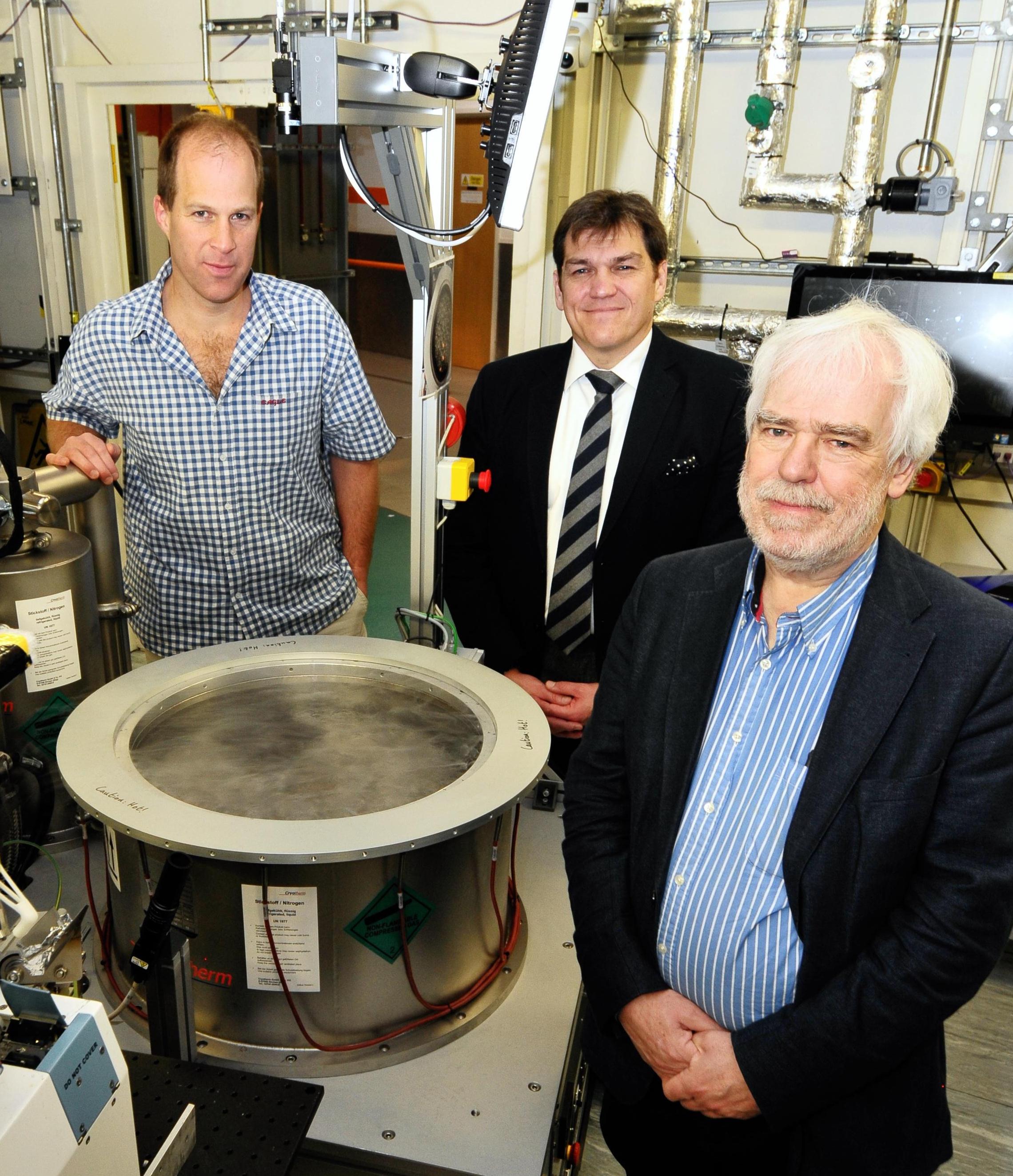
Peter Moody (L), Russell Wallis (M) and Peter Andrew (R)
A recent discovery that has the potential to benefit from the fragment screening facility has been led by a team of scientists from the University of Leicester. They have successfully solved the structure of the elusive protein pneumolysin. This toxin, which is secreted by Streptococcus pneumoniae bacteria, is capable of killing cells. In doing so, it helps these bacteria, which are responsible for a range of serious illnesses including pneumonia, meningitis and septicaemia, to take hold in humans.
Funded by the Wellcome Trust and the Medical Research Council, the University of Leicester research was led by Professor Russell Wallis of the Departments of Infection, Immunity and Inflammation and Molecular and Cell Biology and Professor Peter Andrew, Head of Department of Infection, Immunity and Inflammation. The crystal structure, published in the journal
Scientific Reports, also represents a significant milestone for Diamond. Pneumolysin is the 3,000
th protein structure to be solved at the facility and deposited in the
Worldwide Protein Data Bank since Diamond came online in 2007.
Russell Wallis said: “Our research is about a toxin called pneumolysin produced by a bacterium called pneumococcus(aka Streptococcus pneumoniae). Pneumococcal infections are the leading cause of bacterial pneumonia as well as the cause of a range of other life-threatening diseases such as meningitis and septicaemia. Pneumolysin is instrumental in the ability of pneumococcus to cause disease. The World Health Organization (WHO) estimated that more than 1.6 million people die every year from pneumococcal infections, including more than 800,000 children under 5 years old.
“The aim of the research was to find out how pneumolysin kills our cells, thereby causing tissue damage and contributing to disease. In particular we wanted to find out how multiple copies of the toxin assemble on the surface of cells.
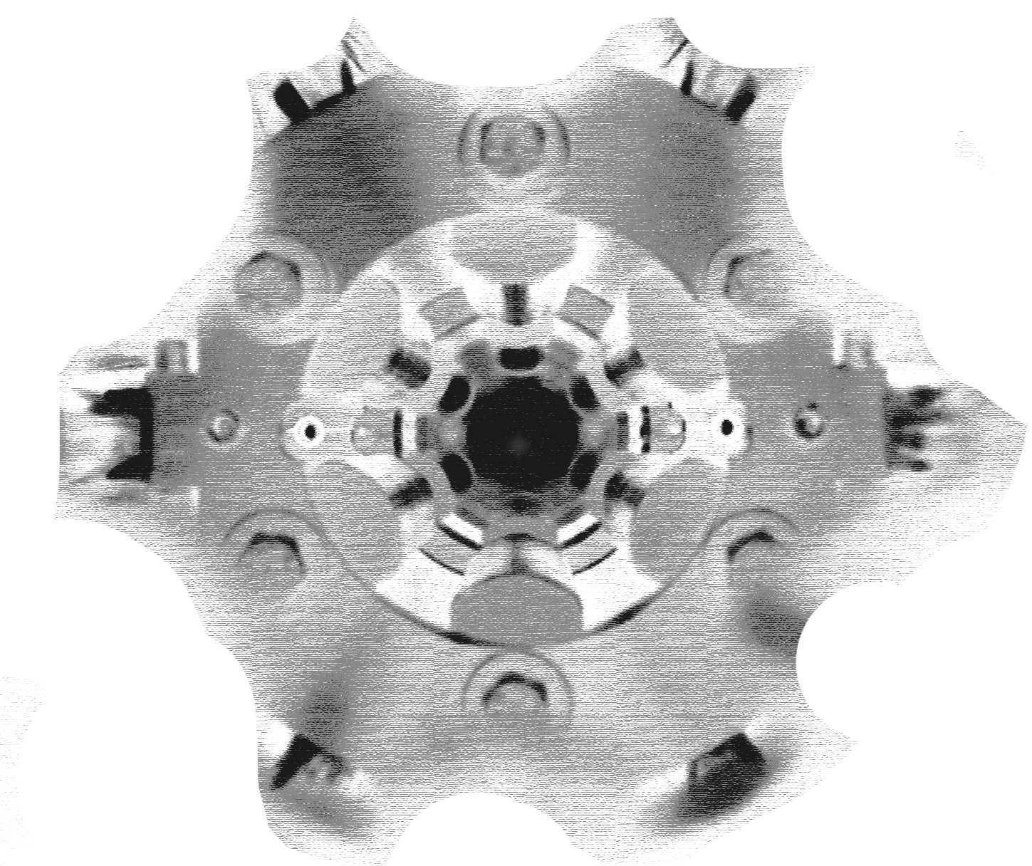
“We managed to determine the structure of pneumolysin using a technique called X-ray crystallography, which enables us to see the individual atoms of the toxin. The structure not only reveals what the toxin looks like, but also shows how it assembles to form lethal pores.
“Ours is the first detailed structure of pneumolysin. This level of detail is important and useful because it enables us to begin to understand how the toxin works. For example, we can see which parts of the toxin come together during pore assembly. When we disrupt these contacts, the toxin becomes inactivated so it can no longer kill cells.
“Using crystallography beamlines at Diamond Light Source, our team has been able to determine the full-length pneumolysin at high-resolution (ten millionths of a millimetre). Down at this level, we can determine the molecular interactions on the cell membrane and work out exactly how the protein forms the pores that are lethal to the body’s cells. Having this knowledge is very exciting as it forms the basis for rational approaches to designing drugs that block assembly of pneumolysin pores to treat people with pneumococcal disease. The University of Leicester has recently set up a company, Axendos Therapeutics, to pursue this aim.
Dave Stuart adds: “This is a fantastic achievement for the Leicester group and a significant milestone for Diamond. We are one of the top synchrotrons in the world when it comes to the number of proteins that scientists are depositing in the Protein Data Bank. We have reached the 3,000th structure at a very exciting point in our development with several new life science beamlines under construction as part of Diamond’s Phase III expansion.”
He continues: “Ultimately, this work reinforces Diamond’s role as a vital source of structural information and demonstrates how integral science is throughout the development process, from fundamental work to delivering new drugs.”


 A brighter light for science