Diamond Home Page
  • Diamond Home Page
  • About Us
  • For Users
  • Industry
  • Public
  • Science
  • Instruments
  • Careers
  • More
Search

On this website

About Diamond

  • About Diamond
  • About Synchrotron
  • News and Features
  • Events
  • Contact Us
  • FAQ

For Users

  • Apply for beamtime
  • User guide
  • Diamond Users Commitee
  • FAQ

Industry

  • Techniques Available
  • Industry Research
  • Industry Case Studies
  • News
  • Meet the Industry Team

Science

  • Research
  • Computing
  • The Machine
  • Publications
  • Research Expertise
  • Membrane Protein Laboratory
  • Additional Facilities

Instruments

  • Biological Cryo-Imaging
  • Crystallography
  • Imaging and Microscopy
  • Macromolecular Crystallography
  • Magnetic Materials
  • Soft Condensed Matter
  • Spectroscopy
  • Structures and Surfaces

Careers

  • Vacancies
  • Info for applicants
  • Company Benefits
  • Apprenticeships
  • PhD Studentships
  • Work Placement

Public

  • News
  • How Diamond Works
  • Multimedia
  • Features
  • Visits Us
  • For School

Procurement

  • Non-OJEU Tender Notices
  • OJEU PINs
  • OJEU Tender Notices
  • Registration Form

Software & Tools

  • Publications Database
  • iSpyB
  • User Administration System

Main Content

Industrial Liaison Office
Sub-navigation
  • Industry Services
  • Our Services
  • Techniques Available
  • Industry Research
  • Case Studies
  • Industry News
  • Meet the Team

In This Section

Sub Navigation
  • News from our Users
  • Latest News
    • 2024

Want to know more?


Enquiry  Make an enquiry

Mailing list  Join our mailing list

Meet the team  Meet the team

____________________________________

Industrial Liaison Group:

Tel: +44 (0) 1235 778797

E-mail: [email protected]

X iconLinkedIn icon

Tweets by @DiamondILO
  1. Industry Services
  2. Industry News
  3. Latest News
  4. Structural Biology Newsletter 6
  5. PanDDA

PanDDA

Electron density for a ligand bound to bromodomain BRD1.

Scientists have utilised Diamond Light Source to develop a new method to extract previously hidden information from the X-ray diffraction data that are measured when resolving the three-dimensional (3D) atomic structures of proteins and other biological molecules.

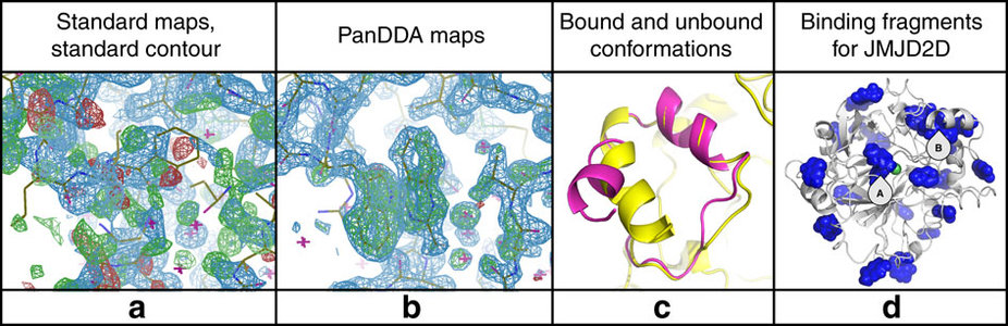
When trying to evolve chemical compounds towards potent drug candidates, scientists attempt to study the atomic detail of how compounds bind to their target proteins. To do so, they compare X ray data measured in both presence and absence of the compound. However, with existing analysis algorithms, this difference signal can often be swamped by noise from experiment artefacts, making it very unreliable to interpret the observed signal.

The new Pan-Dataset Density Analysis (PanDDA) method extracts the picture of the bound compound in exceptionally clear and unambiguous detail. PanDDA first identifies the source of the noise, and then removes it from the data. It exploits Diamond’s ability to repeat dozens to hundreds of measurements quickly, which are then characterised for differences between them, indicating the presence of bound compound, after which a noise correction is applied in 3D. This powerful algorithm is now an integral part of Diamond’s XChem platform and is routinely used on fragment screening campaigns to rapidly extract fragment hits.

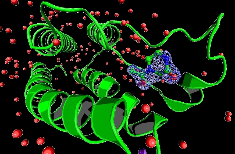
The results have been recently published in Nature Communications. Macromolecular crystallography (MX), the technique that PanDDA applies to, is one of the most powerful tools used by researchers interested in determining the 3D structures of large biological molecules, including proteins, and is the work-horse experiment for rational drug design.

“The problem of identifying binding events in crystallographic datasets can feel like looking for a needle in a haystack,” explains Dr Nicholas Pearce, lead author on the paper which comes from his PhD project at the University of Oxford in the Systems Approaches for Biomedical Science (SABS) Centre for Doctoral Training, where he was jointly funded by UCB Pharma and Diamond. “In the case of the data we were analysing, it was even worse, because we had hundreds of haystacks, and didn’t know which of them contained needles.” Dr Pearce is now based in the Crystal & Structural Chemistry Group at Universiteit Utrecht.
The researchers were able to use to their advantage the fact that most of the measurements were from ‘empty’ crystals that didn’t contain a bound ligand, allowing them to characterise the unbound form and simply looking for datasets that were different.
“Often in crystallography you can miss ‘weak’ bound forms, because each measurement is a superposition of the bound and unbound forms,” continues Dr Pearce. “This is akin to multiple sheets of tracing paper, each with one of at least two images, all overlaid on top of each other.”
“When trying to identify the image on only one of the ‘sheets’, it gets confused by what shows through from all the other sheets, so the image becomes susceptible to interpretation errors,” Dr Pearce adds. “To overcome this, we developed a method to extract the right set of ‘sheets’ from the superposition; once we’d done that, interpreting the bound form becomes much easier, and enables us to confidently interpret the data, and build models of the interesting states in the data.”
“The basic idea is conceptually very simple, namely treating the confusing superposition as a background correction problem,” explains Professor Frank von Delft, who is jointly Principal Investigator of the Protein Crystallography group in the Structural Genomics Consortium (SGC) at the University of Oxford, and Principal Beamline Scientist of the I04-1 beamline at Diamond. “However, an accurate estimate of the background is crucial, and in practice this was unthinkable until the advent of the new robotic technology offered by Diamond, which makes it routine to make such large numbers of measurements.”

“UCB is delighted to have been working closely with Diamond on the development of PanDDA and its application to crystallographic fragment screening,” comments Dr Neil Weir, Senior Vice President of Discovery at UCB Pharma. “As a direct result, we have been able to identify fragments, which were otherwise not distinguishable from background, bound to a key protein-protein interaction drug target.” 

Fig 2: PanDDA maps reveal complex minor conformations and identify allosteric binders (Image © Nature Publications)
The research involved producing around 860 datasets, of which only 75 contain a bound form of interest to the researchers. “While general applicable in MX, the method is particularly transformative for a version of the MX experiment called fragments screening, where the effects we’re looking for are very rare and even harder to verify by conventional algorithms,” continues von Delft.
A crucial coda to the work was the uploading of all the structures to the Protein Data Bank (wwPDB), the online repository of 3D structures of proteins and nucleic acids, where everybody has completely free access to all structures ever published. One of the wwPDB host sites, RCSB PDB, recently developed a new group deposition tool to allow the mass upload of structures, and this was crucial to completing this collaboration.
The RCSB PDB Group Deposition system allows authors to take advantage of local templates and PDB_extract for batch processing, packing, upload, review, validation, and one-click submission of many structures at once. Searching group title “PanDDA analysis group deposition” at rcsb.org will return these 860 depositions.
The researchers hope that this new method will provide a significant shift in how crystallographic models are generated; opening windows to explore more poorly ordered crystals.  

The Industrial Liaison Team at Diamond

Would you like to know more about PanDDA and how you can apply it to your research? Then please get in touch with the Industrial Liaison Team at Diamond. 

The Industrial Liaison team at Diamond is a group of professional, experienced scientists with a diverse range of expertise, dedicated to helping scientists and researchers from industry access the facilities at Diamond. We’re all specialists in different techniques and have a diverse range of backgrounds so we’re able to provide a multi-disciplinary approach to solving your research problems.

We offer services ranging from full service; a bespoke experimental design, data collection, data analysis and reporting service through to providing facilities for you to conduct your own experiments. We’re always happy to discuss any enquiries or talk about ways in which access to Diamond’s facilities may be beneficial to your business so please do give us a call on 01235 778797 or send us an e-mail. You can keep in touch with the latest development by following us on Twitter @DiamondILO or LinkedIn

Pearce, N. M. et al. A multi-crystal method for extracting obscured crystallographic states from conventionally uninterpretable electron density. Nat. Commun. 8, 15123 doi: 10.1038/ncomms15123 (2017), is available at: www.nature.com/articles/ncomms15123.

  • Contact Us
  • About Diamond Light Source
  • Careers
  • Procurement
  • Legal notices & Cookie policy
  • Supply Chain Transparency

Diamond Light Source

Diamond Light Source is the UK's national synchrotron science facility, located at the Harwell Science and Innovation Campus in Oxfordshire.

Copyright © Diamond Light Source


Diamond Light Source Ltd
Diamond House
Harwell Science & Innovation Campus
Didcot
Oxfordshire
OX11 0DE

See on Google Maps

Diamond Light Source® and the Diamond logo are registered trademarks of Diamond Light Source Ltd

Registered in England and Wales at Diamond House, Harwell Science and Innovation Campus, Didcot, Oxfordshire, OX11 0DE, United Kingdom. Company number: 4375679. VAT number: 287 461 957. Economic Operators Registration and Identification (EORI) number: GB287461957003.

feedback