
___________________________________
Industrial Liaison Group:
Tel: +44 (0) 1235 778797
E-mail: industry@diamond.ac.uk
Imaging is defined as "the process of producing an exact picture of something". In the case of synchrotrons, imaging isn't just about seeing what is on the outside of an object, but what is inside of it. Whether it's metals, engineering components, rocks, biological samples or even delicate archaeological samples, X-ray imaging allow us to view what is contained within the sample, at a molecular level.
X-ray imaging allows detailed information to be gathered from below the surface of a material through either full-field imaging, where the whole sample is illuminated, or through scanning, where the beam is focused to a small spot which is scanned across the sample. The high intensity and energy of the synchrotron X-rays we produce here at Diamond make it possible to image a much larger range of materials and sample thicknesses than conventional X-ray sources, and the brilliance of the synchrotron source produces very high resolution images.
These high intensity X-rays also permit very fast measurements for high speed imaging experiments which allow you to look at changes in your sample during real-time, in situ experiments. A technique called X-ray computed tomography can create three dimensional reconstructions of the internal sample volume which makes it possible to view any cross-section of the virtual image at any angle.
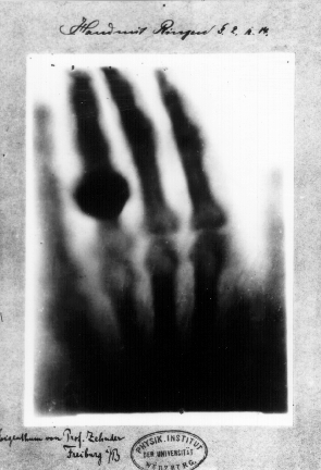
So how did this technique come about? Well, in 1895 German physicist Wilhelm Röntgen was in his laboratory at the University of Munich researching cathode rays – the phenomena where an electrical current passing through an evacuated glass tube causes the end of the tube to glow (or fluoresce) - when he noticed that a screen painted with barium platinocyanide over a metre away on his lab bench was shimmering. Intrigued, he placed a number of objects between the vacuum tube and the screen and still the screen glimmered. Röntgen had discovered X-rays, for which he went on to win the inaugural Nobel Prize for Physics in 1901.
Röntgen's discovery attracted the attention of medical researchers, given the ability of the new "rays" to penetrate beneath the skin, but also physicists who were more interested in trying to establish the nature of the mysterious rays. In 1912 Max von Laue produced the first diffraction pattern and William Lawrence Bragg reformulated von Laue’s conditions for diffraction into what became known as Bragg’s Law, which gives a direct relationship between the crystal structure and its diffraction pattern. X-ray Crystallography was born.
Not long afterwards in Manchester, Ernest Rutherford, alongside his work to split the atom, realised that accelerated particles provided a useful tool for investigating the structure of matter. Physicists around the world began looking into ways to accelerate particles and it was through this work that an alternative source of X-rays grew. The first accelerators (cyclotrons) were built by particle physicists in the 1930s. In these machines, the nucleus of the atom was split using the collision of high-energy particles. From the results of these collisions the physicists tried to deduce the laws of fundamental physics that govern our world and the whole of the universe.
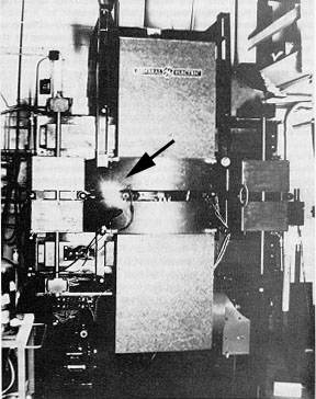
Synchrotron radiation was seen for the first time at General Electric in the United States in 1947. Herb Pollock, Robert Langmuir, Frank Elder, and Anatole Gurewitsch saw a gleam of bluish-white light emerging from the transparent vacuum tube of their new 70MeV electron synchrotron at General Electric's Research Laboratory, Schenectady, New York: Synchrotron radiation had been seen.
It was first considered a nuisance because it caused the particles to lose energy, but by 1956 it was was recognised as light with exceptional properties that overcame the shortcomings of X-ray tubes.
In 1956, the first experiments were carried out using synchrotron light siphoned off from a particle collider at Cornell in the USA. Over the years, the number of experiments using synchrotron light increased, but the scientists still had to use the light that was a by-product of particle collider machines; there was no dedicated synchrotron light source.
In the mid- to late 1970s, scientists began to discuss ideas for using synchrotrons to produce extremely bright X-rays. These discussions led to the construction in the late 1980s and early 1990s of the 'third generation synchrotrons' we now use today. Impressive progress continues to be made in accelerator physics, electronics and computing as well as in magnet and vacuum technologies and there are now almost 50 synchrotron light sources around the world.
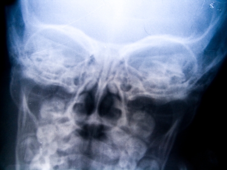
The most common imaging technique is called absorption contrast imaging and it is the technique used in hospital X-ray imaging that we are most familiar with. An absorption contrast image is essentially a shadowgraph, the contrast being generated by the different attenuating power of materials in the sample. The small spot size and high intensity of synchrotron X-rays make is possible to carry out absorption contrast imaging in much finer detail than from conventional sources. Combining absorption contrast imaging with other X-rays techniques allows more detailed complementary information to be gathered, as in diffraction enhanced imaging, or to recreate three dimensional objects from two-dimensional scans as in tomography.
![]()
X-ray tomography is the construction of a three dimensional image from two dimensional projections taken at different orientations. High energy synchrotron X-rays can penetrate through thicker materials, providing a tool for examining the internal features of a sample in a non-destructive way. The parallel, monochromatic beam enhances the image quality beyond what is possible with laboratory techniques.
Tomography has many applications in the material science, engineering and biomedical fields. It can be used to characterise the internal structure of porous materials such as trabecular bone or metal foams. Tomography can be used to determine the size and shape of cracks and other defects inside components such as aircraft and automotive parts, where unexpected failures could have catastrophic results. Because it is non-destructive, X-ray tomography can be used to study the internal structure of precious and unique objects in archaeology and palaeontology – for example studying ancient insects fossilised in amber.
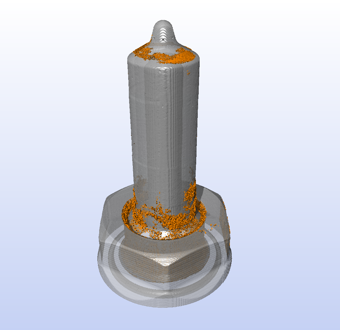
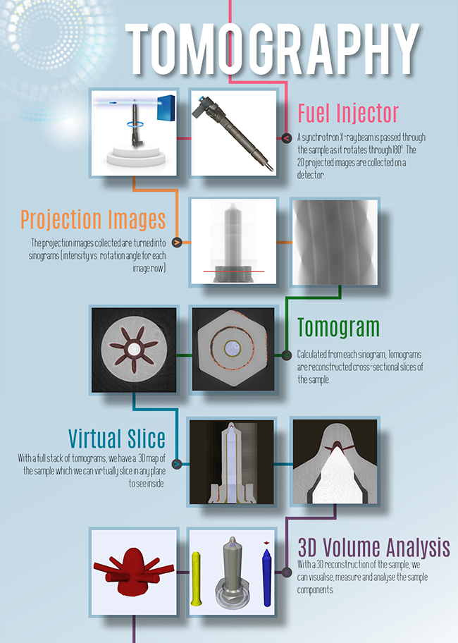
In the diagram below, a fuel injector was imaged at Diamond. The X-ray Beam was passed over the sample as it rotated through 180o and 2 dimensional images were collected on a detector. Each of these 2D projection images was then turned into what is known as a Sinogram ( a plot of intensity vs. rotation angle) and it is these sinograms that allow researchers to calculate a Tomogram - a reconstructed cross-sectional slice of the sample.
This is where the 3D picture really starts to build. With a full stack of tomograms, you have a 3D map of the imaged sample, allowing you slice into and view the inside of the sample in any plane. With the 3D reconstruction of the fuel injector, we are able to visualise, measure and analyse the sample components.
Would you like to know more about imaging and Tomography and how you can apply it to your research? Do you perhaps have a structural problem that you are unable to solve in your lab or a material you wish to find out more about? Then please get in touch with the Industrial Liaison Team at Diamond.
The Industrial Liaison team at Diamond is a group of professional, experienced scientists with a diverse range of expertise, dedicated to helping scientists and researchers from industry access the facilities at Diamond. We’re all specialists in different techniques and have a diverse range of backgrounds so we’re able to provide a multi-disciplinary approach to solving your research problems. We offer services ranging from full service; a bespoke experimental design, data collection, data analysis and reporting service through to providing facilities for you to conduct your own experiments.
We’re always happy to discuss any enquiries or talk about ways in which access to Diamond’s facilities may be beneficial to your business so please do give us a call on 01235 778797 or send us an e-mail. You can keep in touch with the latest development by following us on Twitter @DiamondILO or LinkedIn

Diamond Light Source is the UK's national synchrotron science facility, located at the Harwell Science and Innovation Campus in Oxfordshire.
Copyright © 2022 Diamond Light Source
Diamond Light Source Ltd
Diamond House
Harwell Science & Innovation Campus
Didcot
Oxfordshire
OX11 0DE
Diamond Light Source® and the Diamond logo are registered trademarks of Diamond Light Source Ltd
Registered in England and Wales at Diamond House, Harwell Science and Innovation Campus, Didcot, Oxfordshire, OX11 0DE, United Kingdom. Company number: 4375679. VAT number: 287 461 957. Economic Operators Registration and Identification (EORI) number: GB287461957003.

 Industrial Liaison Office