
Selected presentation abstracts, with an informal introduction from the speakers.

Complex internal structure of dinosaur teeth revealed using synchrotron transmission X-ray microscopy
National Synchrotron Radiation Research Centre
Taiwan Light Source, BL01B1
The question of dinosaur dietary preferences is an interesting issue for paleontologists. Traditionally, analysis of tooth shape and hip structure has been used to identify what different species preferred to eat. In this work, we demonstrated how to use the very modern synchrotron imaging technique, transmission X-ray microscopy (TXM), at BL01B1 of TLS in Taiwan, to observe the internal structure and configuration of various dinosaur teeth with very high spatial resolution. We found out that the internal structure and configuration, as well as the shape of the teeth, strongly correlate to functions of the teeth that tell us much about their dietary preferences. For example, the internal structure and configuration of the long and sharp teeth of a T. rex also exhibit internal features which protect them from damage. Such teeth were useful for hunting prey with a very high damage threshold. The TXM observation shows that there is a relatively soft tissue, called mantle dentin, distributed between enamel and dentin in teeth which has many nano pores inside. This design is very similar to man-made break water stones along the shoreline, which can diffuse the force of waves before they reach the land. This porous mantle dentin disappeared from the crown of plant-eating ornithischian dinosaur teeth when they began to be used for grinding tough plant fibers.
Some plant-eating dinosaurs, however, had long and sharp teeth like the T. rex which could not easily be used for grinding plants, and were only used for raking the plants into their mouths. The plants were actually ground by the many small stones they swallowed, much like a chicken. However, it was less efficient when dinosaurs used the small stones they swallowed to digest the plants than when they used their teeth to do so. Perhaps that is the reason why the plant-eating dinosaurs which had long and sharp teeth went extinct before the final extinction of the dinosaurs, because they could not compete with the dinosaurs who had grinding teeth to obtain food. By using the TXM, we discovered a possible evolution story behind the fossil teeth that can be traced back several hundred million years.
Abstract
COMPLEX INTERNAL STRUCTURE OF DINOSAUR TEETH REVEALED USING SYNCHROTRON TRANSMISSION X-RAY MICROSCOPY
C.-C. Wang1*, C.-C. Chiang1, R. R. Reisz2, and Y.-F. Song1
1. 101 Hsin-Ann Road, Hsinchu Science Park, Hsinchu 30076, Taiwan, R.O.C.
2. Department of Biology, University of Toronto at Mississauga, Mississauga, ON L5L 1C6, Canada.
Tooth protection mechanism of animals with high biting force has long attracted great scientific interest. Amazingly the maximum biting forces of extinct Tryrannosaurus rex and extant crocodiles are typically higher than tens of thousands Newtons. What causes the material design of such high fracture resistant teeth? In this study, we applied synchrotron transmission X-ray microscopy (TXM) at Taiwan Light Source [1] to identify the complex 3D internal ultramicrostructures of various kinds of dinosaur and crocodile teeth within a phylogenetic framework (see Fig. 1)[2]. We found that the internal microstructures are very different between saurischian and advanced ornithischian teeth, reflecting differences in dental developmental strategies and mechanical functions.
From TXM, we found relative soft mantle dentin with tiny interglobular porous space exhibits near the dentinoenamel junction (DEJ) in saurischian teeth represents the primitive condition of dinosaur teeth. Mantle dentin, greatly reduced or absent from DEJ in derived ornithischian teeth, is a key difference between Saurischia and Ornithischia. The stress shielding and crack resistance protective functions of mantle dentin with interglobular porous microstructures inside typical saurischian teeth are discussed using finite-element analysis method [2]. Recent progress will also be presented.
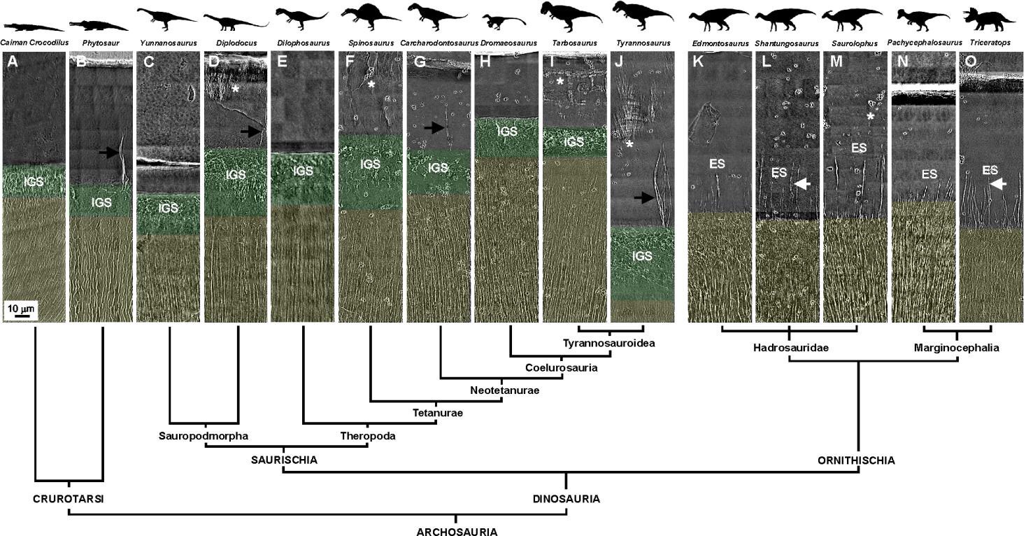
Figure 1: Internal tooth microstructures of various dinosaur genera within a comparative phylogenetic framework: Asterisks indicate enamel cracks. Black and white arrows indicate enamel tufts (ET) and the periodic features of a long enamel spindle (LES), respectively. Here, IGS: interglobular porous space structure; and ES: enamel spindle. Mantle dentin and dentin are colored translucent green and yellow, respectively. Enamel is uncolored.
References
- Yin, G C, et. al., “Energy-tunable transmission x-ray microscope for differential contrast imaging with near 60 nm resolution tomography,” Appl. Phys. Lett. 88, 241115 (2006).
- Wang, C C, et. al., “Evolution and Function of Dinosaur Teeth at Ultramicrostructural Level Revealed Using Synchrotron Transmission X-ray Microscopy,” Sci. Rep. 5, 15202 (2015).
Chun-Chieh Wang
 Chun-Chieh works at BL01B1 beamline of TLS in Taiwan, and is interested in studying the internal structure and configuration of organs and tissues of extinct animals and insects through fossils. He wants to find out their mechanical and biological functions and learn more about the evolution of their structural features by using the TXM. He is also interested in developing some new 3D image registration and reconstruction algorithms for the imaging processing of the transmission X-ray microscopy and the X-ray micro-computed tomography.

Diamond Light Source is the UK's national synchrotron science facility, located at the Harwell Science and Innovation Campus in Oxfordshire.
Copyright © 2022 Diamond Light Source
Diamond Light Source® and the Diamond logo are registered trademarks of Diamond Light Source Ltd
Registered in England and Wales at Diamond House, Harwell Science and Innovation Campus, Didcot, Oxfordshire, OX11 0DE, United Kingdom. Company number: 4375679. VAT number: 287 461 957. Economic Operators Registration and Identification (EORI) number: GB287461957003.