
Keep up to date with the latest research and developments from Diamond. Sign up for news on our scientific output, facility updates and plans for the future.
The Materials Village beamlines provide a variety of experimental techniques for studying a diverse range of materials. The four village beamlines: Materials and Magnetism beamline (I16), Small-Molecule Single-Crystal Diffraction beamline (I19), Test beamline (B16) and the X-ray Imaging and Coherence beamline (I13) - with one branchline operated in collaboration with the University of Manchester - continue to produce exciting new science. In addition to actively supporting a diverse user programme, the village beamlines continue to develop new equipment and techniques to maintain their state-of-the-art.
While I16 accommodates a broad range of science, from Grazing Incidence – Small Angle X-ray Scattering (GI-SAXS) to fundamental X-ray physics, its raison d’etre is to carry out studies of magnetic and electronic ordering phenomena using resonant single-crystal diffraction. Such projects account for the majority of the beamline use including for example the article ‘Unveiling the magnetic ordering mechanism in Ru based hexagonal perovskites’ in this Annual Review. In that work, a number of advanced experimental and theoretical techniques were brought to bear and solve a highly complex magnetic structure.
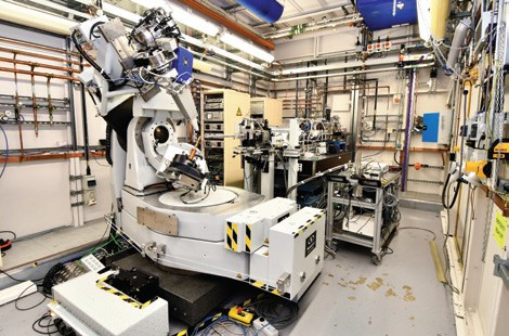
Figure 2: I16’s six-axis kappa diffractometer in its experimental hutch.
In January 2007, the beamline celebrated the 10th anniversary of its -and Diamond’s- first users. Ten years on and both the scientific ‘hot topics’ and experimental techniques have changed enormously. Both of these have dictated the evolution and development of the beamline, such as an extension of the energy range down to the ruthenium L-edges, and taking advantage of the latest in area detector technology. Two new projects – a microdiffraction stage and an advanced polarization analyser, with an in vacuum area detector – are at an advanced stage and expected to be available for users in the coming months. Coupled with other planned developments, the beamline should maintain its position as a key research tool in the rapidly evolving field of condensed matter physics, for the UK and international science communities, throughout the next decade.
After seven years of user operation, I19 had a significant upgrade in 2015 with the installation of a new diffractometer. The new instrument allows users to collect data from smaller, more weakly diffracting, crystals than was previously possible, with a more efficient and faster data collection. The new instrument was designed by a team of Diamond’s engineers and the I19 beamline scientists, and some aspects of its design borrowed heavily from similar installations on Diamond’s macromolecular crystallography (MX) beamlines.
The new diffractometer has three-circle geometry, incorporating a unique dual air-bearing system, which allows the sample to be positioned and rotated extremely accurately, so that very small crystals, less than 5 microns in size, can be reoriented while remaining accurately centred within a tightly focused X-ray beam. The incorporation of a photon-counting Pilatus 2M detector allows data collection in ‘shutterless’ mode where diffraction data, in the form of 2D images, are read continuously, while the diffractometer rotates the sample at a constant speed in the beam. This data collection method is much more efficient than before and a data collection can now be performed in approximately five minutes rather than the former 90 minutes.
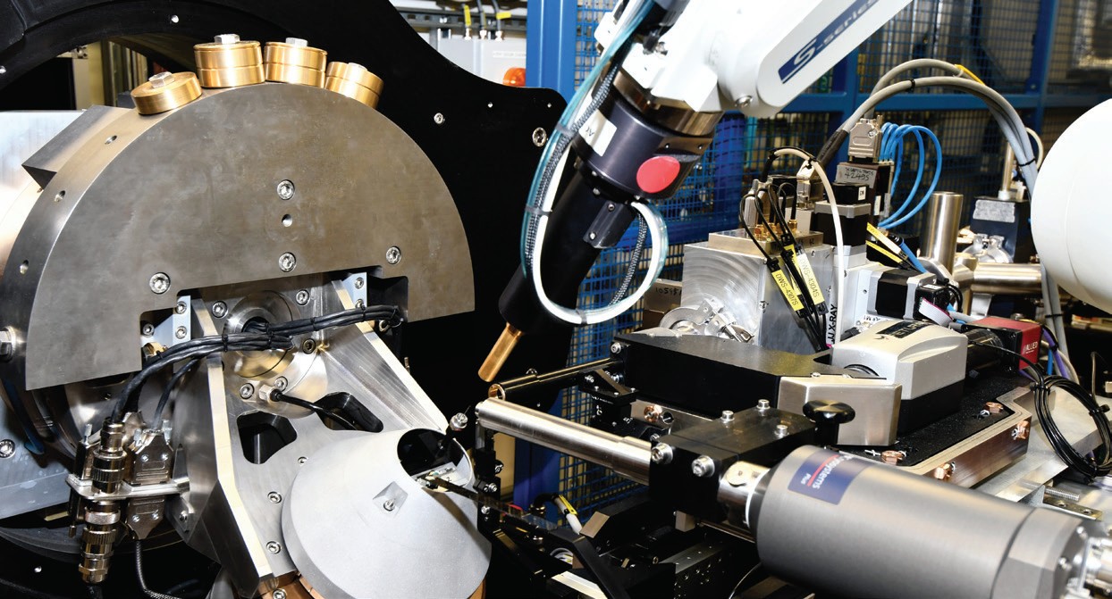
Figure 1: The goniometer and diffractometer of I19
Accompanying the significant improvement in the data collection efficiency there has been a substantial increase in the throughput of samples for chemical crystallography on the beamline. The higher throughput places greater demands on sample preparation to minimise pauses between data collections while the next sample is selected and placed on the diffractometer. Therefore, a means of storing a group of samples and mounting them in an automated manner, without the need to enter the hutch, is necessary to make best use of beamtime. The use of the robotic sample changer will, therefore, become an essential element to the efficient operation of the new end station. At the end of 2016 the existing robot was upgraded to the new BART system so that both it and the diffractometer are fully integrated into the beamline controls infrastructure.
The storage and transportation of samples is a key element in the use of the robot and the beamline has now adopted the methodologies developed for MX for storing a large number of pre-mounted samples under liquid nitrogen and transporting them to Diamond in a Dewar. Once at Diamond the samples are loaded into the Dewar of the robot, while still being held under liquid nitrogen, so that the robot can then mount the samples on to the diffractometer. With the samples now stored and ready for data collection within experimental hutch, there is no need for users to re-enter the hutch during their beamtime. The members of the North East England BAG were the first to exploit this new mode of operation where, after sending their samples by courier to Diamond, they controlled the beamtime remotely from Newcastle.
The 250 m long Diamond beamline I13 is dedicated to high-resolution imaging, coherence diffraction imaging (CDI) and tomography; and operates two independent branchlines. The imaging branchline, also called the Diamond-Manchester branch, performs micro-tomography. Customised sample environments and rigs are implemented on the setup, catering for a large variety of experiments and user communities. The areas of interest are very diverse such as studying structure and properties of bones, the vision and navigation of insects, materials under impact, strain and stress, to name just a few. Regarding beamline instrumentation, most recently significant progress has been made in the implementation of a full-field microscope. It will provide sub-100 nm resolution with exposure times of several hundred milliseconds. It can operate in Zernike-mode for weakly absorbing specimens. The instrument will accommodate sample environments developed and procured with the Diamond-Manchester collaboration. The recently installed multilayer monochromator allows element-specific imaging with short exposure times (some 100 ms).
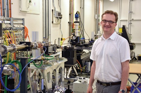
Figure 3: Principal Beamline Scientist, Christoph Rau, in one of the experimental hutches of I13.
The collaboration with Manchester University has provided a large variety of science for bio-medical applications, batteries, materials science and even studying ice cream. The relationship has developed further and will be expanded in the near future. We expect in particular jointly to develop sample environments.
The coherence branch is producing high impact science, for example imaging chromosomes with a Multilayer Laue Lens (MLL)-based microscope, providing a 13 nm x 33 nm focal spot (see article ’Viewing chromosomes with X-rays on the nanoscale’ in this Review). With ptychographic scanning 30 nm structures are resolved; tomographic scans can be done in combination with fluorescence measurements. The main progress here is the improved user-friendliness of the software and with it the user support provided. The second main method on the coherence branch is Bragg-CDI. The detectorsupporting robot arm is now fully implemented and software commissioned. The EXCALIBUR detector provides photon counting over a large 3 megapixel array and has been recently commissioned for user operation.
During the last year the I13-data ‘beamline’ has become operational. The facility allows users to return to Diamond for data analyses, using the Diamond infrastructure and state-of-the-art computing capabilities. The visits can be booked through the UAS; a room at I13 is available for data analyses and support is provided by beamline staff.
The Test Beamline (B16) is designed to provide a highly flexible experimental environment. The diversity of equipment options available on the beamline allow us to accommodate the uncertainties associated with the trial of new experiments. The beamline routinely provides support to other beamlines and technical groups within Diamond in order to characterise new optics, detectors and instrumentation. The flexibility of the schedule permits a rapid response to such requests. The external user community frequently work with the beamline to perform experiments ranging from transmission and fluorescence imaging of biological samples to diffraction from novel engineering materials. Many of these experiments utilise the versatility of the beamline to facilitate new experimental developments. Upgrades to the beamline in 2016 have included high speed piezo stages for raster-fluorescence scanning and significantly improved specimen illumination allowing improved microscope images of samples to be recorded during experiments.
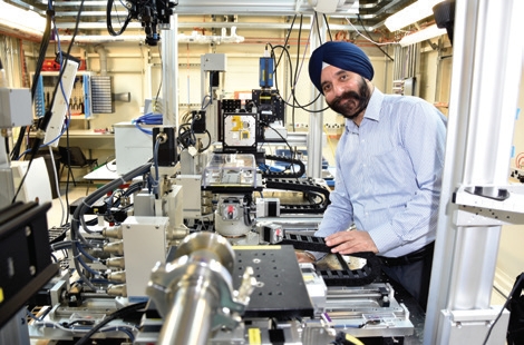
Figure 4: Principal Beamline Scientist, Kawal Sawhney with the versatile experimental and sample platform of B16.

Diamond Light Source is the UK's national synchrotron science facility, located at the Harwell Science and Innovation Campus in Oxfordshire.
Copyright © 2022 Diamond Light Source
Diamond Light Source Ltd
Diamond House
Harwell Science & Innovation Campus
Didcot
Oxfordshire
OX11 0DE
Diamond Light Source® and the Diamond logo are registered trademarks of Diamond Light Source Ltd
Registered in England and Wales at Diamond House, Harwell Science and Innovation Campus, Didcot, Oxfordshire, OX11 0DE, United Kingdom. Company number: 4375679. VAT number: 287 461 957. Economic Operators Registration and Identification (EORI) number: GB287461957003.

 A brighter light for science