
Keep up to date with the latest research and developments from Diamond. Sign up for news on our scientific output, facility updates and plans for the future.
Kawal Sawhney, Village Coordinator
The Materials Village beamlines provide a variety of experimental techniques for studying a diverse range of materials. The four village beamlines: Materials and Magnetism (I16), Small-Molecule Diffraction (I19), the Test beamline (B16) and the Imaging and Coherence beamline (I13, with one branchline operated in collaboration with the University of Manchester) continue to produce exciting new science. In addition to actively supporting a diverse user programme, the village beamlines continue to develop new equipment and techniques to maintain their state-of-the-art facilities.
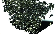 Data gathered at the Diamond Manchester Imaging Branchline (I13-2) have been used to gain a better understanding of lithium battery failure. Lithium-ion batteries provide high power and energy densities, with cells containing pure lithium electrodes theoretically offfering the highest energy storage density by weight and volume. However, lithium metal is unsuitable for commercial use in batteries because it is considered inherently unsafe. Repeated charging and discharging results in electrodeposition of lithium into tree-like dendritic deposits, termed ‘lithium moss’. These deposits continually grow and can short-circuit the battery, leading to battery failure and potentially to fires or explosions.
 Anisotropic materials are those that exhibit chemical or mechanical differences depending on their orientation. A common example of such a material is wood, where the direction of the grain has an impact on the wood’s characteristics. Birefringent materials are optically anisotropic: the refractive index of the material is dependent on the polarisation state of light passing through it. So, by observing the effect on polarised light passing through a birefringent sample, we can establish the nature of the anisotropy within it. Using the polarising optical microscope, this idea has been used extensively over the last century and has had significant impact on many scientific disciplines including mineralogy, crystallography, materials science, and biological sciences.
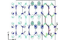 The understanding of magnetism and its effects is important for the fundamental understanding of materials, as well as having the potential to lead to important technological advances. To understand many magnetic effects one needs to study deep at the atomic level, as it is the collective behaviour of all the individual magnetic moments of the constituent atoms that determines the observed macroscopic magnetic properties. At high temperatures magnetic materials are paramagnetic, with the directions of the magnetic moments (spins) randomised by thermal fluctuations. However, at low temperatures magnetic moments tend to point in a particular pattern to minimise the magnetic interactions.
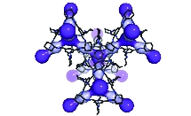 Rare gases exist at very low concentrations in the Earth’s atmosphere; with the exception of argon, noble gases such as xenon and krypton are found at less than two parts per million by volume. It is, however, commercially beneficial to extract such gases due to their value; for example, in commercial lighting, medical imaging and anaesthesia in the case of xenon. In contrast, certain isotopes of these gases can present an environmental hazard. Radon occurs in a naturally radioactive form, which can accumulate in buildings and is a leading cause of lung cancer. It is very difficult to separate such molecules at very low concentrations in the air. It is also difficult to separate chiral molecules in solution, where mirror images of the same molecule have an identical size and shape. Ground-breaking research into porous organic cage molecules has shown selective binding of both noble gases and chiral organic molecules.

Unlike the conventional absorption imaging, both phase and dark field contrast imaging cannot be measured directly. Even though a few imaging techniques have been developed over the last few decades, many of them are limited by either sophisticated experimental conditions or stringent beam properties. We have developed a novel imaging technique to extract the dark field imaging from a stack of speckle images1. The new method can also provide directional dark-field information, which is extremely useful for the study of strongly ordered systems.
In order to reveal the internal structure of soft tissues, the speckle based phase contrast imaging has also been extended from 2D radiography to 3D tomography2. The initial work was carried out on B16, and further experiments were performed on PETRA’s P05 beamline to produce the first 3D image of a human artery by using speckle based technique. As shown in Fig.2, significant enhanced contrast has been observed in the phase contrast computed tomography (CT). The simplicity of the experimental arrangement and speed of measurement gives this new imaging method a distinct advantage over existing X-ray imaging methods, and as such, makes it an attractive technique for in vivo imaging of soft tissue biological systems.

References
1. Wang H. et al. Hard-X-Ray Directional Dark-Field Imaging Using the Speckle Scanning Technique. Phys. Rev. Lett. 114, 103901 (2015).
2. Wang H. X-ray phase contrast tomography by tracking near field speckle. Scientific Reports. 5, 8762 (2015).
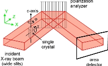
Here, we demonstrate that by careful choice of a single, highly sensitive Bragg reflection, it is possible to map the surface of an extended crystal with intensity contrast of more than two orders of magnitude, by adopting a pair of photon energies just above and below an absorption edge. Such a map can be carried out with microfocusing optics to achieve micron-scale (or better) spatial resolution. We illustrate this technique with a study of artificially-poled domains in KTiOPO4 1 – a ferroelectric material that exhibits important nonlinear optical properties.
The sensitivity of various Bragg reflections to photon energy, near the Ti K edge, was first investigated numerically using the CCTBX crystallographic library, with Diamond’s DAWN data analysis package as a Python programming platform. These simulations predicted a huge contrast from several reflections, exceeding a factor of 200 for the (417) Bragg reflection.
Measurements were carried out on I16 using the Beamline’s Kirkpatrich- Baez (KB) microfocusing mirror system, giving a spatial resolution, after accounting for beam footprint effects, of around 1.5 x 1.7 microns. The (417) Bragg reflection was measured, with the beam rastered over the sample, for two photon energies, just above and below the Ti K-edge. Thus the ‘domain fraction’ could be determined from the intensity ratio, eliminating any effects of sample inhomogeneity. The resulting map (Fig. 1) shows large regions of up ‘A’ domains towards the top with ‘B’ domains dominating the majority of the scanned area. Towards the centre, a set of artificially generated inversion domains (vertical lines) can be seen, with a periodicity of ~ 9 microns. These were created artificially by applying a high voltage through special electrodes2 in order to improve optical phase-matching for frequency-doubling applications. The results show a very clear map of the artificial domain pattern, and indicated that domain reversal is far from complete, reaching only about 40% at most, with regions where the domain inversion failed completely.
Reference:
1. F Fabrizi et al. Acta. Cryst. A (submitted for publication).
2. T Lyford et al. Acta. Cryst. A (accepted for publication).
Beamline I13 consists of two independently operating branchlines for imaging and coherence. Micro- and nano- structures are imaged in the energy range of 6-35keV, making use of the coherence of light. The beamline covers different scientific areas such as biomedicine, materials science and geophysics.

Diamond Light Source is the UK's national synchrotron science facility, located at the Harwell Science and Innovation Campus in Oxfordshire.
Copyright © 2022 Diamond Light Source
Diamond Light Source Ltd
Diamond House
Harwell Science & Innovation Campus
Didcot
Oxfordshire
OX11 0DE
Diamond Light Source® and the Diamond logo are registered trademarks of Diamond Light Source Ltd
Registered in England and Wales at Diamond House, Harwell Science and Innovation Campus, Didcot, Oxfordshire, OX11 0DE, United Kingdom. Company number: 4375679. VAT number: 287 461 957. Economic Operators Registration and Identification (EORI) number: GB287461957003.

 A brighter light for science